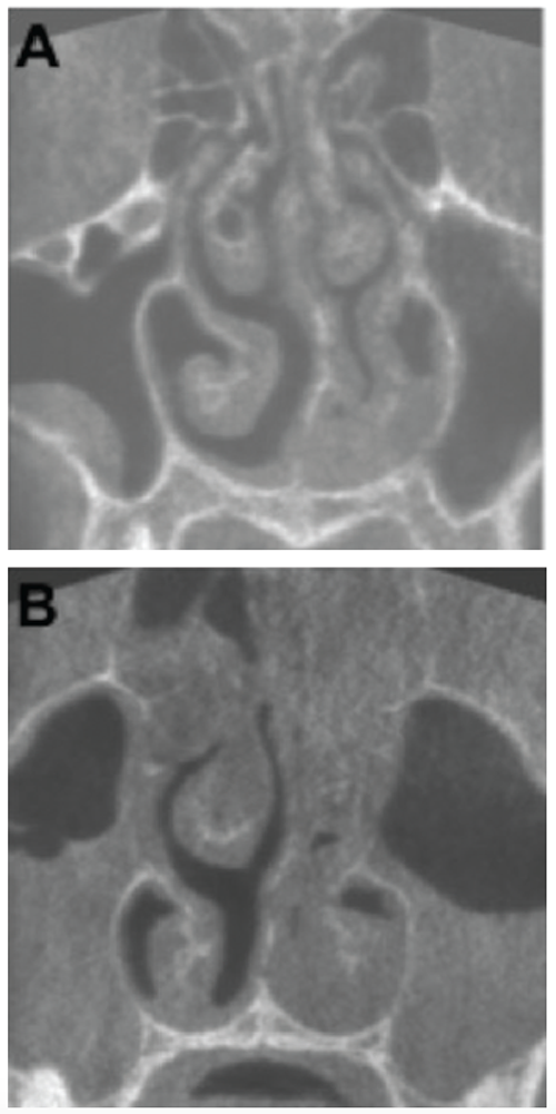
KA) Coronal plane of a CBCT scan from a patient three days after onset of symptoms. Haller cell and concha bullosa are present on the right side, and a cyst is present in the right maxillary sinus. The nasal septum is deviated to the left (19.4°). Both osteomeatal complexes are open, and other paranasal sinus mucosal abnormalities are not present. (B) Coronal plane of a CBCT scan from a patient nine days after onset of symptoms. There are air-liquid levels in the left maxillary sinus and gas bubbles in the right. The patient underwent maxillary sinus aspiration, which was cultured for Haemophilus influenzae.