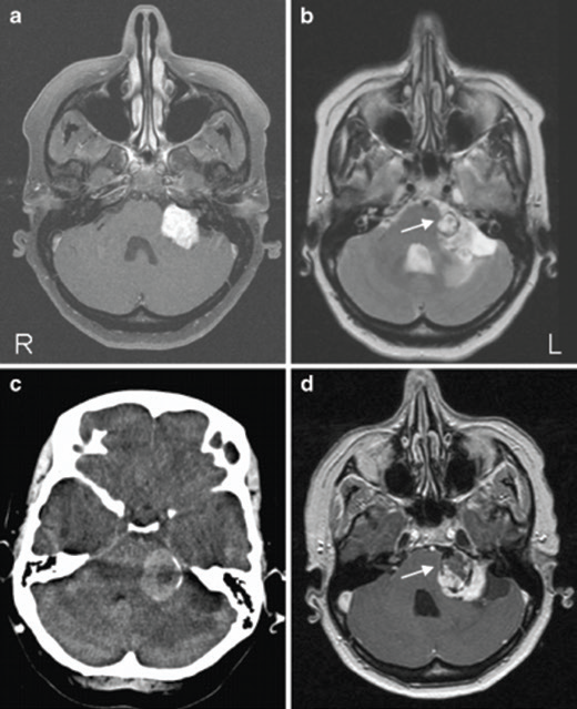
Caption: A) Axial T1-weighted MRI with gadolinium contrast enhancement showing left-sided vestibular schwannoma (VS). B) Axial T2-weighted MRI of the posterior fossa showing round lesion inside the VS (arrow) with surrounding hemosiderin deposits. C) Axial CT image through the posterior fossa showing a hyperdens area due to intratumoral hemorrhage in the VS. D) Axial T1-weighted postcontrast MRI showing hemorrhage (hypo-intense area) inside lesion (arrow). The VS enhances homogeneous and contrast-sparing occurs at the location of the hematoma (R right, L left).