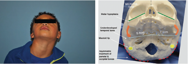
Figure 1. Left: preoperative image demonstrating degree of hemifacial microsomia highlighted by deficient mastoid and asymmetric midface and mandible. Right: 3D-printed skull highlighting these changes at the bony level.
Figure 1. Left: preoperative image demonstrating degree of hemifacial microsomia highlighted by deficient mastoid and asymmetric midface and mandible. Right: 3D-printed skull highlighting these changes at the bony level.
ENTtoday - https://www.enttoday.org/article/how-to-3d-customization-for-microtia-repair-in-hemifacial-microsomia/ent_0322_pg12a/