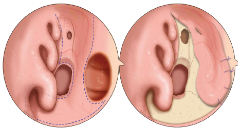
Figure 1. Schematic picture of the extended anterior ethmoidal artery (AEA) flap. (Left) Endoscopic view of the limits of the AEA flap, including the entire mucosa of the nasal floor and inferior meatus flap and the sphenoid rostrum. (Right) Endoscopic view of a large septal perforation completely repaired with the extended AEA flap suturing to the flap edges. The entire nasal floor, inferior meatus, and sphenoid rostrum are left denuded