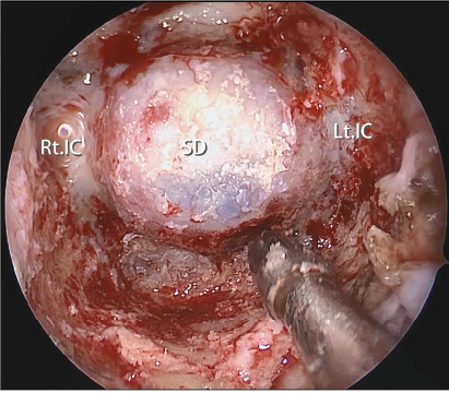
Fig. 1. Final endoscopic view after resection of the anterior wall of the sphenoid sinus including the PVC to the medial edge of the Vidian canal. Note the inferolateral part of the anterior sphenoidotomy is maximized, expanding the working corridor for surgical instruments. SD = sellar dura; Lt. IC = left internal carotid artery; Rt. IC = right internal carotid artery.