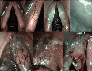
Examples of different Ni classification types applied in studied group of patients. Type I: thin, interconnected vessels. Type II: enlarged vessel diameter. Type III: vascular patterns obscured by white mist. Type IV: small regularly distributed dots. Type Va: solid or hollow thick brown spots with various shapes. Type Vb: destroyed loop, its remnants are visible as line-like shapes. Type Vc: combination of type Va and Vb, irregular distribution.