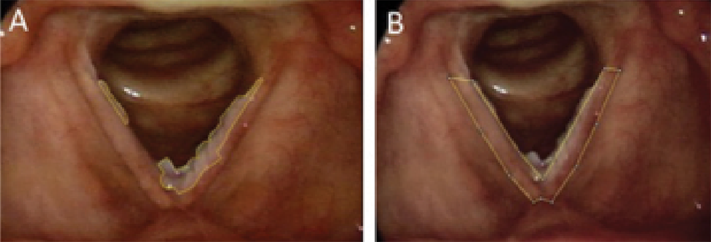
Figure 2. Determination of % lesion regression. (A) Average lesion area (three measurements) = 1,831 pixels. (B) Average full VF area (three measurements) = 6,924 pixels. Percent VF covered by the lesion was determined by
(A/B) × 100% = (1,831/6,924) × 100% = 26.4% of the VF covered by lesion. This % was then compared with the postoperative picture % to determine regression.
Credit: Copyright 2017 The American Laryngological, Rhinological and Otological Society, Inc.