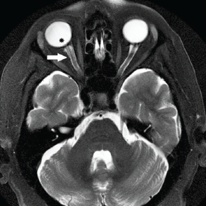
Axial, T2-weighted MRI demonstrating optic nerve sheath dilation (white arrow) and flattening of the posterior aspect of the right globe (asterisk).
Axial, T2-weighted MRI demonstrating optic nerve sheath dilation (white arrow) and flattening of the posterior aspect of the right globe (asterisk).
ENTtoday - https://www.enttoday.org/article/otolaryngologists-play-key-role-in-management-of-idiopathic-intracranial-hypertension/ent_0418_pg10b/