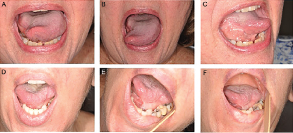
Figure 1. Granulation postoperatively. (A–C) Patient six months after partial glossectomy involving one-third of the tongue and small area of lateral floor of mouth. (D–F) Patient 15 months after partial glossectomy involving one-third of the tongue and lateral floor of mouth. Note adequate tongue protrusion with only mild tethering in both patients
Credit: Copyright 2017 The American Laryngological, Rhinological and Otological Society, Inc.