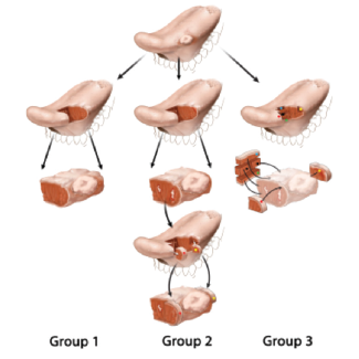
An exophytic tumor at the lateral border of the oral tongue is illustrated. In group 2, white irregular areas represent residual carcinoma at the margin. In groups 2 and 3, colored dots represent tumor bed margins.
An exophytic tumor at the lateral border of the oral tongue is illustrated. In group 2, white irregular areas represent residual carcinoma at the margin. In groups 2 and 3, colored dots represent tumor bed margins.
ENTtoday - https://www.enttoday.org/article/specimen-based-margin-analysis-early-tongue-cancer-better-predict-local-control/ent_0817_pg6a/