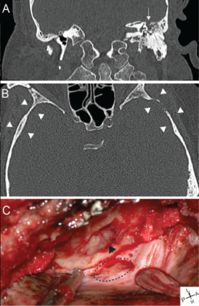
Figure 2: Representative images from a patient with a lateral sCSF leak. (A) Representative coronal CT showing left tegmen mastoideum defect (arrow) with fluid in the middle ear and mastoid. (B) Axial CT demonstrating cortical skull thinning (arrowheads). (C) Intraoperative images showing tegmen defect with encephalocele (arrowhead) and dural defect (dotted line).
Copyright 2017 The American Laryngological, Rhinological and Otological Society, Inc.