 TRIO Best Practice articles are brief, structured reviews designed to provide the busy clinician with a handy outline and reference for day-to-day clinical decision making. The ENTtoday summaries below include the Background and Best Practice sections of the original article. To view the complete Laryngoscope articles free of charge, visit Laryngoscope.
Background
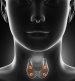
Primary hyperparathyroidism is the most common cause of outpatient hypercalcemia and affects approximately 1% of the population (Ann Surg Oncol. 2011;19:981-989; JAMA Surg. 2016;151:959-968; Ann Surg Oncol. 2012;19:577-583). Unifocal adenomas account for around 85% of disease presentation, with multifocal glandular hyperplasia responsible for a majority of remaining cases (Ann Surg Oncol. 2011;19:981-989; JAMA Surg. 2016;151:959-968; Ann Surg Oncol. 2012;19:577-583). Diagnosis is made biochemically via measurements of total calcium, parathyroid hormone, creatinine, and 25-hydroxyvitamin D levels (JAMA Surg. 2016;151:959-968). Familial hypocalciuric hypercalcemia is assessed preoperatively during a 24-hour urine collection (JAMA Surg. 2016;151:959-968). Surgery is the only definitive treatment for primary hyperparathyroidism and is indicated for patients with symptomatic disease, serum calcium levels >1 mg/dL above normal, cases demonstrating renal involvement, patients with osteoporosis, patients under the age of 50, patients with psychiatric symptoms attributable to hypercalcemia, or patients with coexisting conditions at risk for complications related to hypercalcemia (JAMA Surg. 2016;151:959-968).
CLIPAREA | Custom media / Shutterstock
Neck exploration remains the gold standard when offering surgical treatment for primary hyperparathyroidism. Minimally invasive single-gland exploration can be offered when preoperative imaging identifies a high likelihood of unifocal disease and intraoperative hormone monitoring reveals an adequate response to extirpation. Common preoperative imaging modalities include ultrasound, scintigraphy, computed tomography (CT), and magnetic resonance imaging (MRI). The following review aims to detail available imaging modalities with the goal of identifying an optimal preoperative imaging modality when evaluating a patient with primary hyperparathyroidism.
Best Practice
Ultrasound, scintigraphy, 4D CT, and MRI all demonstrate utility when evaluating a patient with primary hyperparathyroidism. Ultrasound offers a real-time anatomic assessment and high-diagnostic sensitivity, making this the evaluation of choice by many parathyroid surgeons. Four-dimensional CT offers increased diagnostic accuracy and cost efficacy when compared to traditional nuclear medicine evaluations. Despite its recent emergence, additional high-quality evidence may result in coalescence around 4D CT as the modality of choice when assessing patients with primary hyperparathyroidism.