 As a child, I had several ventilation tubes inserted into my left ear, including a long-term tube. I remember having to move from an exam room to a procedure room, lying on a flat, cushioned examination table, and having my ear suctioned. I hated it. When the ventilation tube was eventually removed, I needed a tympanoplasty. After the surgery, my auricle felt numb, I couldn’t hear, and it hurt. I still bear a large (4 cm) horizontal scar over my left ear (temporalis fascia) with a bump where the hair doesn’t grow. Even at the time (late 1980s), it seemed like an awfully large incision. My left ear had a tendency to feel clogged, and it was always harder to equalize.
Explore This Issue
January 2026
In 2015, after returning home from a trip, my left ear clogged during descent and remained so for several days. I knew something was off. At the end of the day, back at work, seeing patients, I put an endoscope in my ear, and I found a small retraction pocket of the pars flaccida, accumulating debris. For goodness’ sake, I had a cholesteatoma!
I followed it for about a year with endoscopic self-examination. I had my medical partner debride my ear under the microscope every few months. He would pull out squamous debris, and the ear would ache, then feel slightly better. I knew I was eventually going to need surgery. I wanted that surgery to be endoscopic.
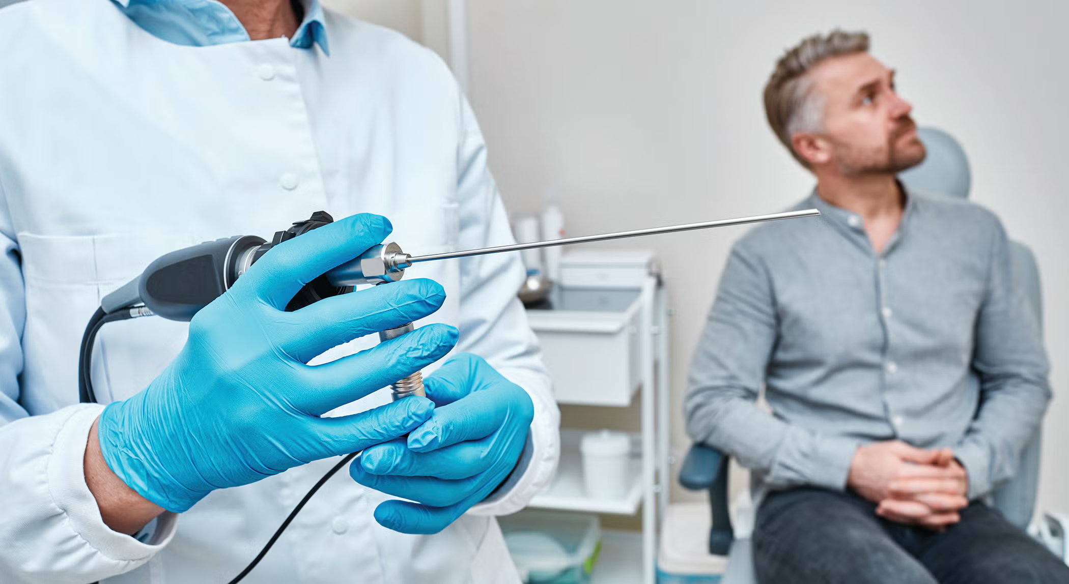
Using endoscopes in ear surgery has always made sense to me. I never used them in residency or fellowship, but during residency, I attended several Academy lectures about endoscopic ear surgery by Muaaz Tarabichi, MD. He was talking about something unique and interesting, and it fascinated me. I admired Dr. Tarabichi for his commitment to developing a new skill and for advocating for it. Those presentations convinced me that endoscopic ear surgery has a place in our specialty.
After I completed an otology fellowship and began practicing, I performed ear surgeries microscopically, which was how I had been trained. However, I added a step to satiate my curiosity and to assess my outcome of the microscopic surgery. At the end of each case, and after I was convinced that I had removed all cholesteatoma, I would ask for an endoscope and tower. I wanted to confirm I wasn’t missing anything, especially in the posterior mesotympanum. I wanted to apply what already made good sense to me; I wanted to see around the corner. The OR staff would sigh (I knew they were thinking I was difficult), but they dutifully did as I asked. I would not infrequently find a small bit of cholesteatoma hidden around a corner when I was certain it had all been removed. So, I wanted to make a change in my surgical practice.
Thank you for sharing your insights!