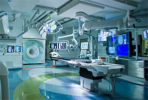
During intra-operative scanning, the MRI travels in and out of the operating room using ceiling-mounted rails to provide real-time images without moving the patient.
Image Credit: Copyright 2015 IMRIS, Inc.
For Joseph Paydarfar, MD, using a CT scan to take images of a patient before transoral surgery has had its limitations.
Like many physicians, Dr. Paydarfar, the section chief of otolaryngology, audiology, and maxillofacial surgery at Dartmouth-Hitchcock Medical Center and associate professor of surgery at the Geisel School of Medicine at Dartmouth College in New Hampshire, relied on the scans taken prior to surgery to see a patient’s anatomy and understand where particular landmarks are located. But when a device is placed in the mouth after surgery begins, the area moves, and the landscape changes, making it difficult to rely on the pre-surgical image as a map. Historically, for neurosurgical applications, patients had their scans taken outside the operating suite and were then moved to the operating room for surgery. If another image was required, the patient had to be returned back to the area where CT scans were taken, adding time to the procedure.
But, with new technology that combines both MRI and CT imaging capability directly in the operating room, doctors like Dr. Paydarfar are now able to use intra-operative imaging, a process by which photos can be taken before, during, and after a surgery if necessary, to show a fuller perspective of the anatomy and the results of a surgical procedure—all while the patient remains in the operating room. With CT scans providing information about bone landmarks and MRI images giving more data about soft tissue in the brain, eyes, and similar types of structures, merging the data that both technologies provide gives a physician far more detail than ever before. Dr. Paydarfar is currently recruiting patients for a research study that will show exactly how the critical structures in the mouth and throat move as surgical devices are placed into the oral cavity. “The goal of this research is be able to predict mathematically how the airway is deformed with the application of various surgical devices,” Dr. Paydarfar said.
 With this research, we’ll be able to get a glimpse of the airway that we haven’t been able to see before, and see how things change. —Joseph Paydarfar, MD
“It’s pretty exciting because, with this research, we’ll be able to get a glimpse of the airway that we haven’t been able to see before, and see how things change,” he said. “We’ll also be able to help expand our capabilities in terms of minimally invasive approaches and making them safer and more effective. But to see how their anatomy is changing—we’ve never been able to visualize this before. It’s really cool stuff.”
Imaging Origins
Physicians began using scans—typically CT scans—prior to surgeries in the early 1990s, said Marvin Fried, MD, one of the first physicians to do so. Dr. Fried, a professor and university chairman in the department of otorhinology–head and neck surgery at the Einstein College of Medicine and Montefiore Medical Center in Bronx, New York, was working at Boston’s Brigham and Women’s Hospital in the mid-1990s, and he used CT scans for endoscopic sinus surgery, along with the first MRI open magnet system, considered a precursor to the MRI machinery now available at Dartmouth and select medical centers around the country. While the use of CT scans for surgery has become more common today, using MRI images before procedures didn’t take hold the same way, he said.
“CT guidance has caught on remarkably well, because it’s easy to set up, and everybody can get a CT scan before sinus surgery,” said Dr. Fried. “It reduces the incidence of adverse events and allows a surgeon to get more accomplished because they know where they are in the anatomy. MRI, however, never really caught on universally because it is very expensive and you need a lot of equipment compatible with a high magnetic field, he said.
CT scans have also been critical for sino-nasal and skull base surgery, said Timothy Smith, MD, MPH, chief of rhinology and sinus surgery, professor of otolaryngology–head and neck surgery at Oregon Health and Science University in Portland, and an ENTtoday editorial advisory board member. “There were early adopters of the technology. but it has found a home even in mainstream otolaryngology,” he said. “I think most surgeons across the country have access to it.”
How Imaging Is Used Now
Combining the CT and MRI technologies is fairly novel—Dartmouth opened its intra-operative imaging center in February 2014—and only 40 other medical centers in the United States, including Brigham and Women’s Hospital in Boston and Barnes-Jewish Hospital in St. Louis, Mo., offer this combination of scanning in the operating room.
At Dartmouth, the first U.S. location to install the machines and receive FDA approval, imaging machinery is suspended on ceiling railings that slide in and out of place in four operating rooms, said Sohail K. Mirza, MD, MPH, medical director of the Center for Surgical Innovation, professor and chair of the department of orthopaedics at Dartmouth’s Geisel School of Medicine. While such technology is still in its earliest stages of use in otolaryngology procedures, it has been used for surgery of the brain, breast, lung, prostate, liver, pancreas, kidneys, and spine.
In these procedures, the extra perspective is helpful. “It’s useful to have a three-dimensional map of where you are and what lies below the surface,” said Dr. Mirza, particularly in cases where the anatomy has a deformity or a congenital malformation. For pediatric spinal cases, where the area is small and patients often don’t have “the normal anatomical landmarks surgeons use for orientation,” the technology has been beneficial, he said.
In a recent spinal case he oversaw, Dr. Mirza described a 75-year-old patient with arthritis-related tumors and curvature of the spine resulting in pinched nerves, spinal stenosis, and numbness/weakness going down the legs. “The patient had already seen five spin surgeons and all recommended a huge surgery to fuse most of the lower back, taking eight to 10 hours. This kind of scoliosis surgery in older adults can take a year to recover from, with a high risk of potentially serious complications,” Dr. Mirza said. “He came to me and said, ‘I am very active. Can you do something to free up my nerves without having to fuse my back?”
Dr. Mirza said he could.
He used the imaging during surgery to locate exactly where the patient’s nerves were, and performed bone removal in six small areas without removing ligaments. “Usually we use indirect measurements with probes to check if the nerves are free when we are finishing up,” he said. “I thought we were done, but when we looked on the CT and MR images after the initial procedure, on one area, one area still looked pinched. We had the ability to fix that.” The entire surgery took four hours, plus an hour for the follow-up CT and MR images, and then another half-hour to fix the final area. As a result, “we were able to remove the minimum amount of bone using the imaging and navigation available, and the patient has been very happy with the results,” he said.
Future Use?
As the technology improves, physicians will likely be able to use intra-operative imaging in more procedures, particularly as research is published showing the results of its use.
Dr. Mirza is eager to see what happens. He cited Dr. Paydarfar’s research on new retractors developed using intra-operative imaging and incorporating validation algorithms that can be developed to determine what kind of pressure is required when using a retractor, and how it affects the look and behavior of a tongue during transoral surgery.
He welcomes other researchers to Dartmouth as well. “We would love to invite projects from anywhere in the country, to build enough of a research core from grants and philanthropy so surgeons, residents, and students who have good ideas to improve surgery don’t have to spend years trying to secure funding, but can come here to do their projects, get their data, and quickly put the good ideas into practice,” said Dr. Mirza. “We also want to work with similar facilities to measure patient outcomes carefully so we can better understand optimal use of this advanced technology in the future.”
Cheryl Alkon is a freelance medical writer based in Massachusetts.