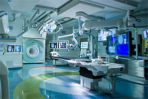
During intra-operative scanning, the MRI travels in and out of the operating room using ceiling-mounted rails to provide real-time images without moving the patient.
Image Credit: Copyright 2015 IMRIS, Inc.
For Joseph Paydarfar, MD, using a CT scan to take images of a patient before transoral surgery has had its limitations.
Explore This Issue
May 2015Like many physicians, Dr. Paydarfar, the section chief of otolaryngology, audiology, and maxillofacial surgery at Dartmouth-Hitchcock Medical Center and associate professor of surgery at the Geisel School of Medicine at Dartmouth College in New Hampshire, relied on the scans taken prior to surgery to see a patient’s anatomy and understand where particular landmarks are located. But when a device is placed in the mouth after surgery begins, the area moves, and the landscape changes, making it difficult to rely on the pre-surgical image as a map. Historically, for neurosurgical applications, patients had their scans taken outside the operating suite and were then moved to the operating room for surgery. If another image was required, the patient had to be returned back to the area where CT scans were taken, adding time to the procedure.
But, with new technology that combines both MRI and CT imaging capability directly in the operating room, doctors like Dr. Paydarfar are now able to use intra-operative imaging, a process by which photos can be taken before, during, and after a surgery if necessary, to show a fuller perspective of the anatomy and the results of a surgical procedure—all while the patient remains in the operating room. With CT scans providing information about bone landmarks and MRI images giving more data about soft tissue in the brain, eyes, and similar types of structures, merging the data that both technologies provide gives a physician far more detail than ever before. Dr. Paydarfar is currently recruiting patients for a research study that will show exactly how the critical structures in the mouth and throat move as surgical devices are placed into the oral cavity. “The goal of this research is be able to predict mathematically how the airway is deformed with the application of various surgical devices,” Dr. Paydarfar said.
 With this research, we’ll be able to get a glimpse of the airway that we haven’t been able to see before, and see how things change. —Joseph Paydarfar, MD
“It’s pretty exciting because, with this research, we’ll be able to get a glimpse of the airway that we haven’t been able to see before, and see how things change,” he said. “We’ll also be able to help expand our capabilities in terms of minimally invasive approaches and making them safer and more effective. But to see how their anatomy is changing—we’ve never been able to visualize this before. It’s really cool stuff.”
Imaging Origins
Physicians began using scans—typically CT scans—prior to surgeries in the early 1990s, said Marvin Fried, MD, one of the first physicians to do so. Dr. Fried, a professor and university chairman in the department of otorhinology–head and neck surgery at the Einstein College of Medicine and Montefiore Medical Center in Bronx, New York, was working at Boston’s Brigham and Women’s Hospital in the mid-1990s, and he used CT scans for endoscopic sinus surgery, along with the first MRI open magnet system, considered a precursor to the MRI machinery now available at Dartmouth and select medical centers around the country. While the use of CT scans for surgery has become more common today, using MRI images before procedures didn’t take hold the same way, he said.