INTRODUCTION
Juvenile nasopharyngeal angiofibromas (JNAs) are rare tumors of the sinonasal cavity that almost exclusively affect adolescent males. JNAs are locally invasive and may extend into and involve adjacent structures. Surgical excision represents the primary treatment; however, the hypervascular nature of JNAs may limit the extent of resection.
Explore This Issue
August 2024Embolization techniques are utilized preoperatively to devascularize tumors and facilitate safer resection, but this remains challenging given the complex neovascular architecture of JNA. There is significant variation regarding embolization practices, with a wide range of techniques described and a limited basis for expert recommendations to guide decision making (Otolaryngol Head Neck Surg. 2023;doi.org/10.1002/ohn.303. Laryngoscope. 2023;doi.org/10.1002/lary.30616.)
Direct puncture embolization (DPE) using an elastic polymer comprising ethylene–vinyl alcohol copolymer, or Onyx (Medtronic, Minneapolis, Minnesota), has been shown to be a safe and effective technique for vascular tumors (Cureus. 2021;13:e15804). As an embolic agent, Onyx offers advantages over n-Butyl-cyanoacrylate (nBCA), including more controlled delivery time, a slower rate of polymerization, and a longer working time (Neuroradiology. 2013;55:1089-1096). In addition, it is more easily visualized under fluoroscopy. Although several small studies have described the use of DPE as an alternative to transarterial embolization (TAE) in treating JNA, management using tandem Onyx DPE with TAE has seldom been described.
METHODS
Institutional review board approval was obtained. Six consecutive male patients aged 14-24 years presented to the same medical center and the same surgeon with primary complaints of nasal congestion and/or epistaxis. Computed tomography (CT), magnetic resonance (MR) radiographic studies, and endoscopic examination were consistent with JNA. All patients were referred to interventional neuroradiology for preoperative embolization followed by endoscopic surgical resection by an otolaryngologist within 24 hours. Age, primary symptoms, tumor staging, tumor size, embolization technique, blood loss, and mean fluoroscopy time were recorded. Tumors were staged according to the Radkowski grading system. The diagnoses were confirmed with histopathology.
TECHNIQUE
On the day prior to surgical resection, patients were positioned within the biplane angiography suite along with endoscopic equipment and appropriate materials to manage nasal hemorrhage.
Step 1: Angiography
Diagnostic angiography was performed, with attention given to the internal carotid artery (ICA). A diagnostic catheter was left in the ipsilateral common carotid artery to perform angiography during the embolization procedure. When angiography demonstrated ICA supply to the JNA (n = 1 patient), a compliant balloon (Eclipse 2L 6 × 20 mm, Balt USA) was appropriately positioned in the distal petrous and proximal cavernous ICA for potential inflation to prevent reflux of the embolic agent into the ICA.
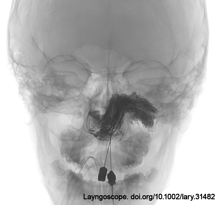
Step 2: DPE
Under fluoroscopic and endoscopic guidance with a 0-degree rigid nasal endoscope, the otolaryngologist advanced dimethylsulfoxide (DMSO)-compatible 20G spinal needles into the mass one at a time to fill each tumor subsite. The number of needles placed depended on the size, shape, and accessibility of the tumor. When the tumor was visible in the posterior oropharynx, needles were also placed transorally. Intratumoral access was confirmed with blood return from the spinal needle. The hub of the spinal needle was then attached to 20-cm DMSO-compatible extension tubing. An intratumoral injection of contrast was performed to identify the extent of vascularity and collateral networks. The tubing and needle were slowly primed with DMSO followed by Onyx-18 embolization under fluoroscopic guidance. Serial assessments of embolization progress were performed with intermittent angiography of the common carotid artery circulation in order to identify residual tumoral blush and progression of embolization material toward dangerous collateral vasculature. Once a particular tumoral compartment had been filled, the same procedure was performed on the next needle. If we determined the need for filling in another part of the tumor, a separate needle was introduced under roadmap guidance. Embolization was continued until no significant tumoral blush was seen on angiography (Fig. 1).
Step 3: TAE
A 5F catheter was navigated into the external carotid artery. Angiography was performed to map the arterial pedicles of the tumor and any residual vascularity. A microcatheter was navigated over a micro guidewire to the distal internal maxillary artery (IMAX). The distal segment of the IMAX was embolized with multiple bare platinum coils to decrease residual blood flow and demarcate the lateral margin of the tumor.
Step 4: Needle Removal
The needles were slowly removed. Onyx was injected slowly during withdrawal to help control bleeding. Minor bleeding from the puncture sites was controlled using direct pressure with oxymetazoline-soaked pledgets under endoscopic guidance. Dissolvable packing was placed to aid in hemostasis. No bleeding required the use of monopolar electrocautery.
A final post-embolization angiogram was performed to determine the degree of tumor devascularization and ensure the patency of intracerebral vessels.
RESULTS
Six consecutive patients with JNA were treated with the tandem DPE + TAE technique between April 2021 and September 2023. Tumors were Radkowsi grade IIa [2], IIb [2], or IIc [2] without extension into the intracranial cavity or orbit. Tumor volume and size varied, with the largest tumor measuring 86 mm in the anteroposterior dimension.
Onyx 18 was preferentially utilized in all patients due to its lower viscosity, which allows the agent to travel more distally and penetrate more deeply than Onyx 34. The average amount of Onyx used for each patient was 16.92 mL (range 6-33 mL). Most cases required one to three needles for injection. The average fluoroscopy time was 60 min. All patients remained neurologically intact after the embolization. There were no complications related to the embolization procedure.
Gross total resection was achieved in all cases. The average estimated blood loss was 335 mL (60-500 mL). The median follow-up time was 209 days (16-757 days). The mean hospital length of stay from the date of embolization to the discharge date postoperatively was 3.7 days (two to six days). All patients underwent MRI imaging on postoperative day one or two to ensure complete removal. Surveillance MRIs were then performed at three to six months post-op and at one year post-op when appropriate. There has been no evidence of recurrent tumors in any patient.