• Home
  • Practice Focus
    • Facial Plastic/Reconstructive
    • Head and Neck
    • Laryngology
    • Otology/Neurotology
    • Pediatric
    • Rhinology
    • Sleep Medicine
    • How I Do It
    • TRIO Best Practices
  • Business of Medicine
    • Health Policy
    • Legal Matters
    • Practice Management
    • Tech Talk
    • AI
  • Literature Reviews
    • Facial Plastic/Reconstructive
    • Head and Neck
    • Laryngology
    • Otology/Neurotology
    • Pediatric
    • Rhinology
    • Sleep Medicine
  • Career
    • Medical Education
    • Professional Development
    • Resident Focus
  • ENT Perspectives
    • ENT Expressions
    • Everyday Ethics
    • From TRIO
    • The Great Debate
    • Letter From the Editor
    • Rx: Wellness
    • The Voice
    • Viewpoint
  • TRIO Resources
    • Triological Society
    • The Laryngoscope
    • Laryngoscope Investigative Otolaryngology
    • TRIO Combined Sections Meetings
    • COSM
    • Related Otolaryngology Events
  • Search

How To: Posterior Septal Artery Flap for Endoscopic Repair of Large Nasal Septal Perforation

by Yury Rusetsky, MD, PhD, Zhanna Mokoyan, MD, Anna Elumeeva, MD, and Olga Chernova, MD • June 17, 2024

  • Tweet
  • Click to email a link to a friend (Opens in new window) Email
Print-Friendly Version

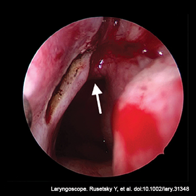
Figure 1. Intraoperative view of the left nasal cavity via a 0° endoscope at the start point of the incision. The arrow indicates the arch of the choana.

To prepare the PSA flap, the posterior incision, made by a bipolar coagulator, started from the posterior end of the middle turbinate (Figure 1), then descended along the posterior edge of the septum obliquely to the border of the hard and soft palate. After that, the incision crossed over the nasal floor and went laterally to the inferior meatus, arising to its lateral wall. Before reaching the lamina of the inferior turbinate, the incision turned anteriorly and went to the axilla of the inferior turbinate. From this point, the incision crossed the nasal floor along the edge of the pyriform aperture, going to the anterior margin of SP. After that, the incision bent around the inferior and posterior edges of the perforation and stopped 5 to 7 mm higher than the posterior border of the perforation.

You Might Also Like

  • How To: Novel Endoscopic Technique to Repair Large Septal Perforation
  • How to: Improving the Outcomes of Anterior Septal Perforations Repair with Combined Flaps
  • Nasal Swell Body Reduction Can Relieve Obstruction in Septal Perforation Repair Patients
  • Can Posterior Septal Nasal Floor Mucosal Flap During Skull Base Reconstruction Repair Cerebrospinal Fluid Leaks?
Explore This Issue
March 2024

It is important to note that, in cases of a combination of “inverted edge” and PSA flap, the medial incision of the PSA flap coincides with the incision for the elevation of the inferior part of the inverted edge flap.

Flap dissection can be performed using a round knife or sharp dissector. Flap elevation started from the pyriform aperture and continued along the nasal floor and the septum to the posterior incision. The most challenging point of dissection was the area of the incisive canal. Hemostasis was achieved by using a monopolar coagulator.

Rotation and placing of the elevated flap were performed using soft tip forceps. The flap should cover the whole perforation without any tension. If the tension persisted, the posterior incision continued to the posterior end of the superior turbinate. According to Zhang et al., at this point, the main stem of the posterior septal artery should be situated. We tried not to make an incision higher than 4 mm from the upper edge of the choana because the distance from the arch of the choana to the inferior branch of the posterior septal artery corresponds to 6.72 6 ± 2.64 mm (Head Neck. 2015. doi:10.1002/hed.23775).

The flap was sutured into the edges of the SP, with single knots at the top, front, and bottom (Figs. 1, 4B), usually using Vicryl 5/0 or Monocryl 5/0. To prevent the formation of dead space, contralateral flaps, and PSA flaps were sutured with several transseptal sutures (Vicryl Rapid 5/0).

At the end of the surgery, thorough hemostasis was achieved through coagulation of the donor site. The inferior nasal meatus was tamponaded with Surgicel. Silicone splints were sutured to the columella for two weeks.

RESULTS

The average operating time was 105 ± 42.5 minutes. In all cases, the highest level of the posterior incision did not run more than 4 mm above the choana edge. It preserves the main stem or inferior branch of PSA (6.72 6 ± 2.64 mm, according to Xian Zhang et al.) (Head Neck. 2015. doi:10.1002/hed.23775). There were no cases of postoperative bleeding or other complications. The wound surface of the nasal floor was completely epithelized one month after surgery.

During the postoperative control visit, complete SP closure was found in 48 of 52 patients (92.3%) in the general group, and in 18 of 22 patients (81.8%) of the “large” SP subgroup, which indicates high effectiveness.   


Pages: 1 2 3 | Single Page

Filed Under: How I Do It, Practice Focus, Rhinology Tagged With: nasal septal perforation, Posterior Septal Artery FlapIssue: March 2024

You Might Also Like:

  • How To: Novel Endoscopic Technique to Repair Large Septal Perforation
  • How to: Improving the Outcomes of Anterior Septal Perforations Repair with Combined Flaps
  • Nasal Swell Body Reduction Can Relieve Obstruction in Septal Perforation Repair Patients
  • Can Posterior Septal Nasal Floor Mucosal Flap During Skull Base Reconstruction Repair Cerebrospinal Fluid Leaks?

The Triological SocietyENTtoday is a publication of The Triological Society.

Polls

Would you choose a concierge physician as your PCP?

View Results

Loading ... Loading ...
  • Polls Archive

Top Articles for Residents

  • Applications Open for Resident Members of ENTtoday Edit Board
  • How To Provide Helpful Feedback To Residents
  • Call for Resident Bowl Questions
  • New Standardized Otolaryngology Curriculum Launching July 1 Should Be Valuable Resource For Physicians Around The World
  • Do Training Programs Give Otolaryngology Residents the Necessary Tools to Do Productive Research?
  • Popular this Week
  • Most Popular
  • Most Recent
    • A Journey Through Pay Inequity: A Physician’s Firsthand Account

    • The Dramatic Rise in Tongue Tie and Lip Tie Treatment

    • Otolaryngologists Are Still Debating the Effectiveness of Tongue Tie Treatment

    • Is Middle Ear Pressure Affected by Continuous Positive Airway Pressure Use?

    • Rating Laryngopharyngeal Reflux Severity: How Do Two Common Instruments Compare?

    • The Dramatic Rise in Tongue Tie and Lip Tie Treatment

    • Rating Laryngopharyngeal Reflux Severity: How Do Two Common Instruments Compare?

    • Is Middle Ear Pressure Affected by Continuous Positive Airway Pressure Use?

    • Otolaryngologists Are Still Debating the Effectiveness of Tongue Tie Treatment

    • Complications for When Physicians Change a Maiden Name

    • Excitement Around Gene Therapy for Hearing Restoration
    • “Small” Acts of Kindness
    • How To: Endoscopic Total Maxillectomy Without Facial Skin Incision
    • Science Communities Must Speak Out When Policies Threaten Health and Safety
    • Observation Most Cost-Effective in Addressing AECRS in Absence of Bacterial Infection

Follow Us

  • Contact Us
  • About Us
  • Advertise
  • The Triological Society
  • The Laryngoscope
  • Laryngoscope Investigative Otolaryngology
  • Privacy Policy
  • Terms of Use
  • Cookies

Wiley

Copyright © 2025 by John Wiley & Sons, Inc. All rights reserved, including rights for text and data mining and training of artificial technologies or similar technologies. ISSN 1559-4939