What features are most significant in diagnosing paresis in patients with vocal fold motion impairment?
Bottom line: Of the multiple features on laryngoscopy, glottic configuration, ipsilateral thin vocal fold, vocal fold bowing, reduced movement, reduced kinesis, and phase lag were more likely to be associated with vocal fold paresis (VFP). There was a relatively poor agreement between the laryngoscopy findings and laryngeal electromyography (LEMG) findings in VFP patients.
Explore This Issue
August 2016Background: The term “paresis” may be defined in many ways, and no fewer than 27 separate features noted on videostroboscopy may indicate VFP. At present, limited data exist to correlate a positive LEMG finding to the variety of endoscopic findings that prompt a clinical paresis diagnosis. If multiple clinical findings that prompt a clinical paresis diagnosis could be simplified and correlated to LEMG findings, paresis diagnosis might be more secure without the need for LEMG.
Study design: Prospective case series of 19 patients suspected of paresis between 2013 and 2014.
Setting: Department of Otolaryngology–Head and Neck Surgery, Department of Neurology, Icahn School of Medicine at Mount Sinai, New York City.
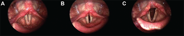
Series of vocal fold images displaying reduced right-sided kinesis. The left vocal fold showed increased excursion with greater movement than the right.
Copyright 2015 The American Laryngological, Rhinological and Otological Society, Inc.
Synopsis: Left-sided VFP was significantly associated with ipsilateral axis deviation, thinner vocal fold, bowing, reduced movement, reduced kinesis, and phase lag. Right-sided VFP was significantly associated with ipsilateral shorter vocal fold, axis deviation, reduced movement, and reduced kinesis. Video strobe laryngoscopy (VSL) findings of ipsilateral axis deviation, reduced movement, and reduced kinesis were significant in both right- and left-sided paresis. For left-sided VFP, a thin ipsilateral vocal fold and lower ipsilateral vocal fold displayed a trend toward statistical significance. For right-sided VFP, ipsilateral phase lag and decreased amplitude approached statistical significance. Of the five stroboscopy measures that did not specify laterality, open phase dominant was the most common finding. Using glottic configuration abnormalities, movement abnormalities, and stroboscopy as key parameters, the senior author was accurately able to diagnose the side of paresis in 89.5% of cases. Endoscopic findings that supported the finding of unilateral VFP on LEMG included axis deviation, bowing, vocal fold thinning, shorter vocal fold, phase lag, and reduced kinesis. Limitations included a limited number of subjects from a single institution and laryngoscopy findings based on two laryngologists with similar training backgrounds.
Citation: Woo P, Parasher AK, Isseroff T, Richards A, Sivak M. Analysis of laryngoscopic features in patients with unilateral vocal fold paresis. Laryngoscope. 2016;126:1831-1836.