• Home
  • Practice Focus
    • Facial Plastic/Reconstructive
    • Head and Neck
    • Laryngology
    • Otology/Neurotology
    • Pediatric
    • Rhinology
    • Sleep Medicine
    • How I Do It
    • TRIO Best Practices
  • Business of Medicine
    • Health Policy
    • Legal Matters
    • Practice Management
    • Tech Talk
    • AI
  • Literature Reviews
    • Facial Plastic/Reconstructive
    • Head and Neck
    • Laryngology
    • Otology/Neurotology
    • Pediatric
    • Rhinology
    • Sleep Medicine
  • Career
    • Medical Education
    • Professional Development
    • Resident Focus
  • ENT Perspectives
    • ENT Expressions
    • Everyday Ethics
    • From TRIO
    • The Great Debate
    • Letter From the Editor
    • Rx: Wellness
    • The Voice
    • Viewpoint
  • TRIO Resources
    • Triological Society
    • The Laryngoscope
    • Laryngoscope Investigative Otolaryngology
    • TRIO Combined Sections Meetings
    • COSM
    • Related Otolaryngology Events
  • Search

A Patient with Right-Sided Odynophagia

by Christine G. Gourin, MD, FACS • February 7, 2011

  • Tweet
  • Click to email a link to a friend (Opens in new window) Email
Print-Friendly Version

Figure 1: Coronal CT scan.

You Might Also Like

  • Patient with Conductive Hearing Loss
  • Dysphagia, Odynophagia, Hoarseness in Elderly Man
  • Psychosocial Aspects of Single-Sided Deafness Important to Consider in Patient Care
  • ‘Doctor, My Ear Hurts’: Diagnosing Referred Otalgia
Explore This Issue
February 2011
Figure 1: Coronal CT scan.

Figure 2: Axial CT scan.

Figure 2: Axial CT scan.

Figure 3: An arrow points to the styloid process.

Figure 3: An arrow points to the styloid process.

Figure 4: The styloid.

Figure 4: The styloid.

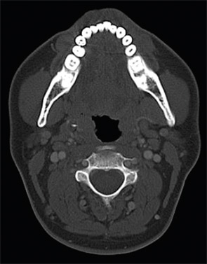
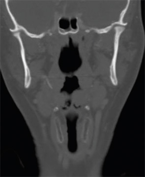
Presentation: A 33-year-old white male presented with a one-year history of right-sided odynophagia. Symptoms were constant and exacerbated by swallowing. He had a history of cryptic tonsils but had not undergone tonsillectomy; his past medical history was otherwise unremarkable. There was tenderness to palpation over the right tonsil with exacerbation of symptoms. No head and neck masses were appreciated. A CT scan was obtained (Figures 1, 2).

What’s your diagnosis? How would you manage this patient? Go to the next page for discussion of this case.

Management: The CT revealed bilateral elongated styloid processes, with the right styloid measuring 4 cm in length and terminating near the right palatine tonsil.

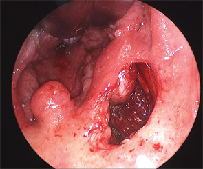
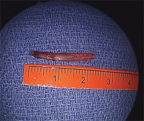
The patient was taken to the operating room, where a palatal incision was made superior to the tonsillar fossa, taking care to avoid injury to the tonsil (Figure 3). It has been the author’s experience that removal of the tonsil does not aid exposure and is not indicated in the absence of tonsillar pathology. Direct palpation of the styloid process is used to guide subsequent dissection, and the tensor palatini muscles overlying the styloid are bluntly elevated off the bone using a Freer elevator. A small rongeur is used to divide the distal 2 cm of the styloid process. At three weeks follow-up, the patient reported complete symptom resolution.

Discussion: Eagle’s syndrome describes a variety of head and neck pain symptoms that may occur secondary to an elongated styloid process. True Eagle’s syndrome is rare; the incidence of an elongated styloid process, defined as greater than 25 mm in length, ranges from 1.4 to 30 percent in the literature, but fewer than 10 percent of patients with an elongated styloid ever present with pain symptoms. Classic Eagle’s syndrome is described as pain from stimulation of cranial nerves IX and X and, at times, V and VII by an elongated styloid. Less commonly, it has been reported that an ossified stylohyoid or stylomandibular ligament may cause cervicalgia or odynophagia that is exacerbated by movement. Glossopharyngeal neuralgia is the most common manifestation of Eagle’s syndrome and the symptom most amenable to relief with a surgical approach when true Eagle’s syndrome is present.

A diagnosis of Eagle’s syndrome is a common referral in otolaryngology practice, but it is important to rule out other causes of pain before attributing symptoms to the styloid process. Because an elongated styloid is a common radiologic finding, confirmation of an association between the styloid process and symptoms may be sought by eliciting symptoms by transoral palpation of the styloid process, or by relieving pain using an injection of topical anesthetic into the tonsil fossa overlying the elongated styloid. If neither maneuver affects symptoms, then it is unlikely that Eagle’s syndrome is present, regardless of radiographic findings.

References

  1. Kim E, Hanson K, Frizzi. Eagle syndrome: case report and review of the literature. Ear Nose Throat J. 2008;87(11):631-633.
  2. Beder E, Ozgursoy OB, Ozgursoy SK. Current diagnosis and transoral surgical treatment of Eagle’s syndrome. J Oral Maxillifac Surg. 2005; 63(12):1742-1745.

—submitted by Christine G. Gourin, MD, FACS, associate professor of otolaryngology-head and neck surgery, Johns Hopkins University, Baltimore, Md.

Pages: 1 2 | Multi-Page

Filed Under: Case of the Month, Otology/Neurotology Tagged With: clinical, OtologyIssue: February 2011

You Might Also Like:

  • Patient with Conductive Hearing Loss
  • Dysphagia, Odynophagia, Hoarseness in Elderly Man
  • Psychosocial Aspects of Single-Sided Deafness Important to Consider in Patient Care
  • ‘Doctor, My Ear Hurts’: Diagnosing Referred Otalgia

Leave a Reply Cancel reply

Your email address will not be published. Required fields are marked *

The Triological SocietyENTtoday is a publication of The Triological Society.

Polls

Have you invented or patented something that betters the field of otolaryngology?

View Results

Loading ... Loading ...
  • Polls Archive

Top Articles for Residents

  • Applications Open for Resident Members of ENTtoday Edit Board
  • How To Provide Helpful Feedback To Residents
  • Call for Resident Bowl Questions
  • New Standardized Otolaryngology Curriculum Launching July 1 Should Be Valuable Resource For Physicians Around The World
  • Do Training Programs Give Otolaryngology Residents the Necessary Tools to Do Productive Research?
  • Popular this Week
  • Most Popular
  • Most Recent
    • Otolaryngologists as Entrepreneurs: Transforming Patient Care And Practice

    • The Dramatic Rise in Tongue Tie and Lip Tie Treatment

    • Continued Discussion And Engagement Are Essential To How Otolaryngologists Are Championing DEI Initiatives In Medicine

    • Rating Laryngopharyngeal Reflux Severity: How Do Two Common Instruments Compare?

    • Otolaryngologists Are Still Debating the Effectiveness of Tongue Tie Treatment

    • The Dramatic Rise in Tongue Tie and Lip Tie Treatment

    • Rating Laryngopharyngeal Reflux Severity: How Do Two Common Instruments Compare?

    • Is Middle Ear Pressure Affected by Continuous Positive Airway Pressure Use?

    • Otolaryngologists Are Still Debating the Effectiveness of Tongue Tie Treatment

    • Complications for When Physicians Change a Maiden Name

    • Physician Handwriting: A Potentially Powerful Healing Tool
    • Leaky Pipes—Time to Focus on Our Foundations
    • You Are Among Friends: The Value Of Being In A Group
    • How To: Full Endoscopic Procedures of Total Parotidectomy
    • How To: Does Intralesional Steroid Injection Effectively Mitigate Vocal Fold Scarring in a Rabbit Model?

Follow Us

  • Contact Us
  • About Us
  • Advertise
  • The Triological Society
  • The Laryngoscope
  • Laryngoscope Investigative Otolaryngology
  • Privacy Policy
  • Terms of Use
  • Cookies

Wiley

Copyright © 2025 by John Wiley & Sons, Inc. All rights reserved, including rights for text and data mining and training of artificial technologies or similar technologies. ISSN 1559-4939