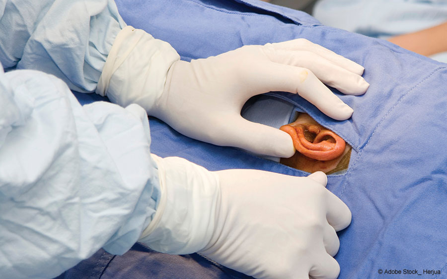
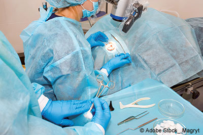
 Scan the literature on transcanal endoscopic ear surgery (TEES) and you’ll find a host of benefits for the procedure when it is compared with its microscope-guided counterpart, including enhanced visualization, superior training, and reduced post-operative complications, to name just a few (Am J Otolaryngol. doi:10.1016/j.amjoto.2020.102451). Coupled with recent equipment advances, such as thinner, more flexible endoscopes and ones that combine cutting and suctioning for enhanced bleeding control, it’s clearly an exciting time for TEES.
Explore This Issue
July 2025It would be hard to find a more dedicated proponent of the technique than Justin S. Golub, MD, MS, vice chair of faculty development in the department of otolaryngology–head and neck surgery at the Columbia University Vagelos College of Physicians and Surgeons and NewYork–Presbyterian/Columbia University Irving Medical Center, in New York City. Dr. Golub is one of several leaders in ENT surgery who are part of a national, concerted effort to drive more widespread adoption of TEES.
A cornerstone of that effort is the American Endoscopic Ear Surgery Study Group (AEESSG), which held its 2024 annual meeting last Fall in Miami, during the American Academy of Otolaryngology–Head and Neck Surgery Foundation (AAO–HNSF) annual conference. Dr. Golub is a board member and past president of AEESSG and part of a panel that presented best practices for TEES treatment of cholesteatomas at the group’s annual meeting. But before offering some highlights from his presentation, he first made a pitch for ENT surgeons with an interest in TEES to join AEESSG.
“This is your chance to be a leader in this field, because it’s still relatively small—fewer than maybe 15% or so of ENT surgeons are performing TEES nationwide,” he said. “It’s an opportunity to help us spread the word and have more champions on board who can speak to and disseminate the advantages of this technique for both the surgeon and the patient.”
Key Benefits of TEES
As for what those key benefits are, Dr. Golub highlighted three he feels make the strongest case for why an ENT surgeon might want to consider TEES. The number one benefit is that the use of minimally invasive surgery avoids having to make a postauricular incision, which is often needed during microscopic-guided tympanoplasty. “That means less pain for the patient, less numbness, and less downtime,” he said.
Those benefits are supported by published data. In a head-to-head comparison of endoscope- and microscope-guided tympanoplasty conducted by Choi et al, pain scores one day after surgery using an 11-item, patient-reported numeric rating scale were significantly less in endoscopically treated patients than in those treated with a microscope-guided approach (Clin Exp Otorhinolaryngol doi:10.21053/ceo.2016.00080). Similarly, a study done by Kakehata et al found that NSAID use was lower for patients treated with TEES (1.3 pills/week) than for those treated with microscopic ear surgery (5.5 pills/week; P<0.001, Mann-Whitney U test). And a study Dr. Golub co-authored cited favorable data for post-operative numbness and other patient-reported outcomes in patients treated via the endoscopic transcanal approach (J Pers Med doi.org/10.3390/jpm12101718).
Perhaps an even more compelling benefit of the endoscope is the superior view of the middle ear that it affords, in part due to its angled orientation, Dr. Golub noted. “You really can see hidden pathology that is often very difficult to visualize with the microscope,” he said. In the case of cholesteatomas, this better visualization is particularly advantageous because it enables the surgeon to see and remove the entire mass more reliably than with a microscope, which in his clinical experience and published data (Int J Pediatr Otolaryngol doi:10.1016/j.ijporl.2020.109872) “results in less recurrences, because you are leaving behind less residual disease,” he said.
The endoscope also reduces the need to perform a canal-wall-down mastoidectomy, “a destructive and invasive” procedure that is sometimes required when using a microscope to better visualize the surgical field, Dr. Golub noted. With the endoscope, in contrast, “you can avoid taking the canal wall down because you can literally just see around it.”
Dr. Golub cited a final key benefit: Endoscopic-guided middle ear surgery “is excellent for training and fosters a sense of camaraderie in the operating room.” With the endoscope, “everyone is looking at the same screen,” he explained. With microscopic ear surgery, in contrast, only the operating surgeon has the ideal view, while everyone else typically views a 2D image on a TV screen “that is often slightly out of focus, sometimes off-center, and may not even show the area the surgeon is working on.”
Kristen Yancey, MD, an assistant professor of otolaryngology-head and neck surgery at Weill Cornell Medicine in New York City and a co-panelist at the AEESSG meeting, echoed and amplified those training benefits. With the shared visualization cited by Dr. Golub, “you’re more able to direct and guide your trainee’s movements and more effectively demonstrate key anatomy,” she said. Moreover, “with an older microscope, the observer’s view can be a little bit darker, which impedes your ability to see certain structures as clearly.”
When working in a small microscopic area, “these subtle differences in lights, optics, and orientation can make a big difference,” Dr. Yancey said.
A Lighter and Better Endoscope
For most evolving surgical techniques, new developments in equipment are key. In the case of EES, Dr. Golub cited the Colibri endoscope (3NT Medical) as an example of such an advancement. Unlike bulky traditional endoscopes, Colibri features “a lightweight ergonomic handpiece, a 2.2-mm diameter tip, and built-in suction to enable two-handed functionality in a single device,” the company noted in a news release announcing the device’s 2020 U.S Food and Drug Administration approval (3NT Medical. https://tinyurl.com/yc6enwtt).
Dr. Golub said he is a fan of the Colibri endoscope for several reasons. “Number one, it’s extremely lightweight,” he said. “It’s made out of hollow plastic, as opposed to traditional endoscopes, which frankly feel like you’re holding a stick with a heavy brick coming out at the end.” Colibri, in contrast, he said, “is light enough so that you can hold it with your non-dominant hand and control its built-in suction with your thumb, while making incisions with the other hand. It’s kind of like operating with one-and-a-half hands, which is a huge improvement over traditional endoscopes.”
Dr. Golub acknowledged that using Colibri is not as good as having two free hands, which is possible during microscope-guided middle ear surgery. “But it’s definitely a huge advancement for TEES.” Asked about Colibri, Dr. Yancey said its built-in suctioning capability “is a great idea. I’m just waiting for the resolution on the camera to get better before I push for it.”
Dr. Golub also stressed that for surgeons interested in learning how to perform TEES, “you don’t need to have the latest and greatest endoscope—you can do this with a 4-mm by 18-cm Hopkins rod-style endoscope that every otolaryngologist has in their operating room for performing sinus surgery.” He added, however, that some tools will make it easier to get started, such as a suction round knife and curved suctions, which are also widely available.
Taking the First Steps
When adding TEES to one’s surgical repertoire, the obvious place to start is education and training, Dr. Golub noted. For that, he reiterated the benefits of joining AEESSG. But as a baseline, “if you’re comfortable using an endoscope for other otolaryngology procedures,” such as the aforementioned sinus surgery, “you have most of the experience you’ll need to start doing [ear] cases.”
Choosing an appropriate first patient, however, is critical, Dr. Golub stressed. “Book a simple case, like ear tubes, and try using the endoscope,” he said. “That’s an easy place to start. Once you’re comfortable, then progress to a simple reconstructive ear surgery, such as a myringoplasty. When you master that, the next step is a tympanoplasty, and it progresses from there.”
Still, patience will be required, Dr. Golub noted. “Even if you are relatively adept at microscopic ear surgery, unless you’ve been trained from the start as an attending at a center where TEES is routinely used, you’re going to be a very slow endoscopic middle ear surgeon—at least in the beginning,” he said. “So it’s really important to give this time, because if you keep at it, you will definitely acquire this skill.”
Once that happens, one of the main payoffs will be speed, Dr. Golub noted. “Ultimately, endoscopic middle ear surgery is faster than most microscopic-guided [procedures] because you don’t have to open a big postauricular incision and then close it,” he reiterated.
That clinical observation is supported by published data. In the Choi et al comparison study, for example, mean operation time for microscopic tympanostomy (MT) was 88.9 + 28.5 minutes longer than that of endoscopic tympanostomy (ET) (68.2+ 22.1 minutes; P=0.002).
Achieving Better Visualization
Once your confidence level with endoscopes in the middle ear builds, it’s useful to start thinking about refining your technique even further, Dr. Yancey noted. During her AEESSG presentation, she focused on one important goal in such efforts—achieving better visualization.
 Positioning the patient is a key first step, Dr. Yancey stressed. She recommended measured use of the reverse Trendelenburg position, which can improve visualization by enhancing venous outflow from the head (Int Forum Allergy Rhinol. doi:10.1002/alr.22734). “I was taught to also have the patient positioned towards the edge of the bed by Dr. Brandon Isaacson, an expert in endoscopic ear surgery at UT Southwestern [Medical Center in Dallas],” Dr. Yancey added. That positioning strategy decreases the “reach” needed to access the ear and therefore increases the surgeon’s stability of maneuvers, she explained.
Moreover, the surgeon’s monitor should be positioned at eye level, directly across from the operative ear, to promote good ergonomics, Dr. Yancey urged. To that end, she recommended using surgical chairs with armrests to support stability while operating. Another useful tip is to set up the operative site so that an instrument wipe and a de-fog pad are readily available. Employing those items “helps efficiently maintain a clear view throughout the case,” she said.
Even basic steps, such as taking time to adequately trim the ear hair and allowing sufficient time for the local anesthetic to take effect, will also help enhance visualization. Otherwise, “you can find yourself fighting blood in the canal and cleaning the lens of your endoscope too frequently,” she said.
Dr. Yancey cited a few additional strategies for controlling bleeding and maintaining a clear line of sight during surgery. In addition to using colloids or pledgets infused with epinephrine for its vasoconstrictive effects (J Otorhinolaryngol Relat Spec. doi:10.1159/000503725), Dr. Yancey recommended using a dedicated suction elevator device that is designed for TEES. “These can be particularly helpful when elevating the tympanomeatal flap, which typically is the bloodiest part of ear surgery cases,” she said. There are a couple of different models designed for this, she noted, including ones from Grace Medical. Whichever device you use, “once you get that initial flap elevated, ear canal bleeding is dramatically improved, and you can really enjoy the benefits of the expanded view that the endoscope affords.”
Leave a Reply