• Home
  • Practice Focus
    • Facial Plastic/Reconstructive
    • Head and Neck
    • Laryngology
    • Otology/Neurotology
    • Pediatric
    • Rhinology
    • Sleep Medicine
    • How I Do It
    • TRIO Best Practices
  • Business of Medicine
    • Health Policy
    • Legal Matters
    • Practice Management
    • Tech Talk
    • AI
  • Literature Reviews
    • Facial Plastic/Reconstructive
    • Head and Neck
    • Laryngology
    • Otology/Neurotology
    • Pediatric
    • Rhinology
    • Sleep Medicine
  • Career
    • Medical Education
    • Professional Development
    • Resident Focus
  • ENT Perspectives
    • ENT Expressions
    • Everyday Ethics
    • From TRIO
    • The Great Debate
    • Letter From the Editor
    • Rx: Wellness
    • The Voice
    • Viewpoint
  • TRIO Resources
    • Triological Society
    • The Laryngoscope
    • Laryngoscope Investigative Otolaryngology
    • TRIO Combined Sections Meetings
    • COSM
    • Related Otolaryngology Events
  • Search

Full-Extension Eyebrow Approach with Supraorbital Nerve Preservation for Frontal Sinus Tumors

by Pedro Plou, MD, Simona Serioli, MD, Alex Y. Alexander, BA, Luciano C. P. C. Leonel, PhD, Maria Peris-Celda, MD, PhD, and Carlos D. Pinheiro-Neto, MD, PhD • January 5, 2024

  • Tweet
  • Click to email a link to a friend (Opens in new window) Email
Print-Friendly Version

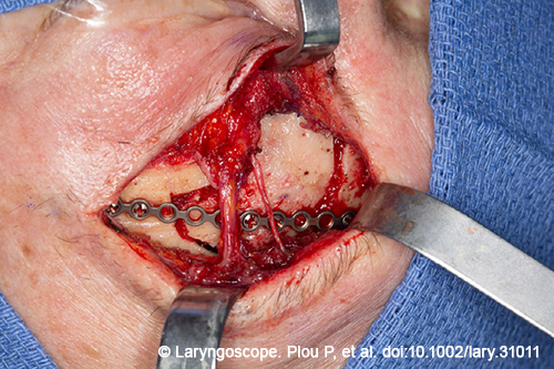
Figure 1. Intraoperative details of the approach and tumor excision. Pictured here is the bone flap reposition and fixation.

An osteoplastic bone flap of the anterior table of the frontal sinus was performed. There was tumor attached to the anterior table of the frontal sinus (bone flap), which was successfully removed. The inner surface of the bone flap was also drilled with a diamond burr to ensure complete tumor resection from the anterior table. The supraorbital nerve was preserved. Complete resection of the tumor was performed, with extirpation of the entire mucosa of the frontal sinus. A high-speed drill with a diamond burr was used to drill out the frontal sinus bony walls and to ensure complete tumor and mucosal extirpation.

You Might Also Like

  • How To: Reconstruction of Anterior Table Frontal Sinus Defects with Pericranial Flap and Titanium Mesh
  • Transorbital Endoscopic Repair a Feasible Approach for Select Patients with Frontal Sinus Cerebrospinal Fluid Leaks
  • How To: Carolyn’s Window Approach to Unilateral Frontal Sinus Surgery
  • Does the Frontal Sinus Need To Be Obliterated Following Fracture with Frontal Sinus Outflow Tract Injury?
Explore This Issue
December 2023

During the cauterization of the posterior table of the FS, there was an area of small dural dehiscence and a cerebrospinal fluid (CSF) leak was observed. Dural synthetic substitute was placed through the opening and immediately stopped the CSF flow. A nasal floor free mucosal graft was also placed to reconstruct the defect. (The management of the intraoperative CSF leak is shown in the intraoperative video). Finally, the anterior table was repositioned and fixed with a titanium plate and microscrews.

No postoperative complications were reported, and the patient was discharged 24 hours after surgery with mild forehead hypoesthesia that recovered completely in three weeks.

RESULTS

The anatomical dissection showed the feasibility of the surgical technique. It allowed enough space for the osteotomy of the entire anterior table of the frontal with preservation of the supraorbital nerve. The lateral, superior, and medial limits of the frontal sinus, including the frontal sinus recess, were well exposed with this approach.

Some anatomical features and extension of the disease may limit the application of an endoscopic eyebrow approach. Frontal sinus anteroposterior dimension below 0.8 cm represents a limitation due to the acute angle and difficult surgical visualization. A pronounced convexity of the posterior wall of the frontal sinus makes it more difficult to manipulate the tumor, reducing intrasinus surgical maneuverability (Ann Otol Rhinol Laryngol. 2012. doi:10.1177/000348941212100802). ]

Moreover, the involvement of the posterior table of the FS entails the risk of CSF leak. As mentioned in the case description, CSF leakage was observed intraoperatively after the removal of the tumor that was successfully repaired through the open approach. Superior located frontal sinus CSF leaks are challenging for endoscopic skull base reconstruction.

In the three cadaveric specimens, the lateral limit of the FS extended lateral to the supraorbital nerve. The degree of exposure was the same as what is obtained with the bicoronal approach. The clinical case illustrated a successful application of this technique.   

Pages: 1 2 3 | Single Page

Filed Under: Head and Neck, How I Do It, Practice Focus Tagged With: Frontal sinus tumorsIssue: December 2023

You Might Also Like:

  • How To: Reconstruction of Anterior Table Frontal Sinus Defects with Pericranial Flap and Titanium Mesh
  • Transorbital Endoscopic Repair a Feasible Approach for Select Patients with Frontal Sinus Cerebrospinal Fluid Leaks
  • How To: Carolyn’s Window Approach to Unilateral Frontal Sinus Surgery
  • Does the Frontal Sinus Need To Be Obliterated Following Fracture with Frontal Sinus Outflow Tract Injury?

The Triological SocietyENTtoday is a publication of The Triological Society.

Polls

Would you choose a concierge physician as your PCP?

View Results

Loading ... Loading ...
  • Polls Archive

Top Articles for Residents

  • Applications Open for Resident Members of ENTtoday Edit Board
  • How To Provide Helpful Feedback To Residents
  • Call for Resident Bowl Questions
  • New Standardized Otolaryngology Curriculum Launching July 1 Should Be Valuable Resource For Physicians Around The World
  • Do Training Programs Give Otolaryngology Residents the Necessary Tools to Do Productive Research?
  • Popular this Week
  • Most Popular
  • Most Recent
    • A Journey Through Pay Inequity: A Physician’s Firsthand Account

    • The Dramatic Rise in Tongue Tie and Lip Tie Treatment

    • Otolaryngologists Are Still Debating the Effectiveness of Tongue Tie Treatment

    • Rating Laryngopharyngeal Reflux Severity: How Do Two Common Instruments Compare?

    • Shifting the Treatment Goalpost Toward Medical Management of Recurrent Respiratory Papillomatosis

    • The Dramatic Rise in Tongue Tie and Lip Tie Treatment

    • Rating Laryngopharyngeal Reflux Severity: How Do Two Common Instruments Compare?

    • Is Middle Ear Pressure Affected by Continuous Positive Airway Pressure Use?

    • Otolaryngologists Are Still Debating the Effectiveness of Tongue Tie Treatment

    • Complications for When Physicians Change a Maiden Name

    • Excitement Around Gene Therapy for Hearing Restoration
    • “Small” Acts of Kindness
    • How To: Endoscopic Total Maxillectomy Without Facial Skin Incision
    • Science Communities Must Speak Out When Policies Threaten Health and Safety
    • Observation Most Cost-Effective in Addressing AECRS in Absence of Bacterial Infection

Follow Us

  • Contact Us
  • About Us
  • Advertise
  • The Triological Society
  • The Laryngoscope
  • Laryngoscope Investigative Otolaryngology
  • Privacy Policy
  • Terms of Use
  • Cookies

Wiley

Copyright © 2025 by John Wiley & Sons, Inc. All rights reserved, including rights for text and data mining and training of artificial technologies or similar technologies. ISSN 1559-4939