• Home
  • Practice Focus
    • Facial Plastic/Reconstructive
    • Head and Neck
    • Laryngology
    • Otology/Neurotology
    • Pediatric
    • Rhinology
    • Sleep Medicine
    • How I Do It
    • TRIO Best Practices
  • Business of Medicine
    • Health Policy
    • Legal Matters
    • Practice Management
    • Tech Talk
    • AI
  • Literature Reviews
    • Facial Plastic/Reconstructive
    • Head and Neck
    • Laryngology
    • Otology/Neurotology
    • Pediatric
    • Rhinology
    • Sleep Medicine
  • Career
    • Medical Education
    • Professional Development
    • Resident Focus
  • ENT Perspectives
    • ENT Expressions
    • Everyday Ethics
    • From TRIO
    • The Great Debate
    • Letter From the Editor
    • Rx: Wellness
    • The Voice
    • Viewpoint
  • TRIO Resources
    • Triological Society
    • The Laryngoscope
    • Laryngoscope Investigative Otolaryngology
    • TRIO Combined Sections Meetings
    • COSM
    • Related Otolaryngology Events
  • Search

How To: A Case Report of Transnasal Endoscopic Drainage for Upper Parapharyngeal Abscess

by John Loh Ming Ren, MBBS, MRCS, MMED, Naomi Huang Wenya, B Pharm, MD, Tan Toh Hui Leonard, MBBS, MRCS, MMED, FAMS, and Pang Cui-Ying Maria, MBBS, MRCS, MMed, FAMS • January 8, 2025

  • Tweet
  • Click to email a link to a friend (Opens in new window) Email
Print-Friendly Version

Figure 1. Image guidance used for incision planning. Salpingopharyngeus muscle and Eustachian tube orifice are visualized.

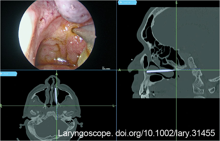
The post-nasal space was examined with a 0-degree Hopkins rod, and the site of the abscess was checked against the image guidance system (Fig. 1). A superficial mucosa biopsy was first performed to exclude nasopharyngeal carcinoma. The biopsy site also served as a marker for the planned incision. A Megadyne 0016 M 6-inch insulated needle point electrode (Ethicon Inc, Cincinnati, Ohio) was used to create a vertical incision medial to the fossa of Rosenmüller, from the roof of the nasopharynx to the level of the Eustachian tube. A vertical incision was preferred over a horizontal incision to avoid lateral dissection toward the Eustachian tube and carotid artery. The salpingopharyngeus muscle served as the lateral landmark to avoid injuring the Eustachian tube orifice (Figure 1).

You Might Also Like

  • New Developments in the Management of Eustachian Tube Dysfunction
  • How To: A Case Report of Transnasal Endoscopic Drainage for Upper Parapharyngeal Abscess
  • Eustachian Tuboplasty: A Potential New Option for Chronic Tube Dysfunction and Patulous Disease
  • Balloon Dilation Is Among the New Frontier of Eustachian Tube Care
Explore This Issue
January 2025

Hemostasis of surface mucosal bleeding was achieved with adrenaline-soaked patties and suction diathermy to ensure good visualization before deeper dissection. Dissection was deepened with a combination of electrocautery dissection and blunt dissection with a Freer elevator, 8 mm suction, and Blakesley forceps. Constant awareness of the depth of dissection was important to prevent inadvertent injury to the carotid artery. This was achieved by measuring the tip length of the instruments as a gauge of depth, and frequent checks against the image guidance system.

Figure 2. Necrotic tissue from the left upper parapharyngeal abscess.

At a depth of 1.3 cm from the mucosal surface, minimal amounts of turbid fluid were encountered posterior–laterally. Blunt dissection was continued toward the source of the turbid fluid till a pocket with necrotic tissue (Fig. 2) was encountered. The necrotic tissue was removed and sent for histological examination and bacteria cultures. The wound was packed with NasoPore (Stryker, Portage, Mich.), which was removed in clinic one week later. The patient was instructed for regular nasal douche.

Aerobic cultures taken from necrotic tissue grew Pseudomonas aeruginosa. Fungal cultures, tuberculosis cultures, and acid-fast bacilli smears were negative. Histology from the superficial biopsy showed mucosa with lymphoid infiltrates with no granuloma or malignancy. Histology of the deep necrotic tissue showed necrotic collagen with neutrophilic aggregates and encrusted bacteria on the surface, with no mucosa or epithelial cells seen. Fluid cultures from the tympanocentesis grew scant Staphylococcus aureus, which was suspected to be contamination from external auditory canal skin flora.

Post-operatively, the patient was kept inpatient for a week of intravenous antibiotic treatment with meropenem, followed by six weeks of oral ciprofloxacin and co-trimoxazole based on antimicrobial culture sensitivities.

CONCLUSION

This study showcases a safe technique for transnasal endoscopic drainage of upper parapharyngeal abscesses. Pre-operative imaging and planning are important to appreciate the relationship to the carotid artery, while intra-operative image guidance and measurement allow the surgeon to maintain anatomical orientation. Taken together, this technique offers effective and safe access for upper parapharyngeal abscess drainage with minimal morbidity and good cosmesis. 

Pages: 1 2 3 | Single Page

Filed Under: Head and Neck, How I Do It, Practice Focus Tagged With: Transnasal Endoscopic Drainage, Upper Parapharyngeal AbscessIssue: January 2025

You Might Also Like:

  • New Developments in the Management of Eustachian Tube Dysfunction
  • How To: A Case Report of Transnasal Endoscopic Drainage for Upper Parapharyngeal Abscess
  • Eustachian Tuboplasty: A Potential New Option for Chronic Tube Dysfunction and Patulous Disease
  • Balloon Dilation Is Among the New Frontier of Eustachian Tube Care

The Triological SocietyENTtoday is a publication of The Triological Society.

Polls

Would you choose a concierge physician as your PCP?

View Results

Loading ... Loading ...
  • Polls Archive

Top Articles for Residents

  • Applications Open for Resident Members of ENTtoday Edit Board
  • How To Provide Helpful Feedback To Residents
  • Call for Resident Bowl Questions
  • New Standardized Otolaryngology Curriculum Launching July 1 Should Be Valuable Resource For Physicians Around The World
  • Do Training Programs Give Otolaryngology Residents the Necessary Tools to Do Productive Research?
  • Popular this Week
  • Most Popular
  • Most Recent
    • A Journey Through Pay Inequity: A Physician’s Firsthand Account

    • The Dramatic Rise in Tongue Tie and Lip Tie Treatment

    • Rating Laryngopharyngeal Reflux Severity: How Do Two Common Instruments Compare?

    • Otolaryngologists Are Still Debating the Effectiveness of Tongue Tie Treatment

    • Is Middle Ear Pressure Affected by Continuous Positive Airway Pressure Use?

    • The Dramatic Rise in Tongue Tie and Lip Tie Treatment

    • Rating Laryngopharyngeal Reflux Severity: How Do Two Common Instruments Compare?

    • Is Middle Ear Pressure Affected by Continuous Positive Airway Pressure Use?

    • Otolaryngologists Are Still Debating the Effectiveness of Tongue Tie Treatment

    • Complications for When Physicians Change a Maiden Name

    • Excitement Around Gene Therapy for Hearing Restoration
    • “Small” Acts of Kindness
    • How To: Endoscopic Total Maxillectomy Without Facial Skin Incision
    • Science Communities Must Speak Out When Policies Threaten Health and Safety
    • Observation Most Cost-Effective in Addressing AECRS in Absence of Bacterial Infection

Follow Us

  • Contact Us
  • About Us
  • Advertise
  • The Triological Society
  • The Laryngoscope
  • Laryngoscope Investigative Otolaryngology
  • Privacy Policy
  • Terms of Use
  • Cookies

Wiley

Copyright © 2025 by John Wiley & Sons, Inc. All rights reserved, including rights for text and data mining and training of artificial technologies or similar technologies. ISSN 1559-4939