• Home
  • Practice Focus
    • Facial Plastic/Reconstructive
    • Head and Neck
    • Laryngology
    • Otology/Neurotology
    • Pediatric
    • Rhinology
    • Sleep Medicine
    • How I Do It
    • TRIO Best Practices
  • Business of Medicine
    • Health Policy
    • Legal Matters
    • Practice Management
    • Tech Talk
    • AI
  • Literature Reviews
    • Facial Plastic/Reconstructive
    • Head and Neck
    • Laryngology
    • Otology/Neurotology
    • Pediatric
    • Rhinology
    • Sleep Medicine
  • Career
    • Medical Education
    • Professional Development
    • Resident Focus
  • ENT Perspectives
    • ENT Expressions
    • Everyday Ethics
    • From TRIO
    • The Great Debate
    • Letter From the Editor
    • Rx: Wellness
    • The Voice
    • Viewpoint
  • TRIO Resources
    • Triological Society
    • The Laryngoscope
    • Laryngoscope Investigative Otolaryngology
    • TRIO Combined Sections Meetings
    • COSM
    • Related Otolaryngology Events
  • Search

How To: Extended External Rhinoplasty Approach for Nasal Dermoids With Intracranial Extension

by Dayton Rand, BA, Brian J. Dlouhy, MD, and Sohit Paul Kanotra, MD • March 15, 2023

  • Tweet
  • Click to email a link to a friend (Opens in new window) Email
Print-Friendly Version

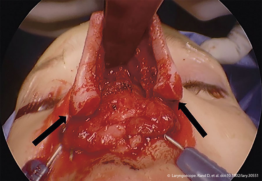
Figure 1. Intraoperative photograph demonstrating the exposure and the alar base extension of the external rhinoplasty incision (black arrows).

An external rhinoplasty approach with an inverted V incision at the mid portion of the columella and marginal incisions was made and extended into alar creases bilaterally. The nasal soft tissue envelope was elevated in a sub-perichondrial plane, and dissection continued over bilateral nasal domes into the lower lateral cartilages. After bisecting the Pitanguay’s ligament, bilateral upper and lateral cartilages were dissected. The incisions were further extended into the alar bases, improving exposure of the nasal dorsum and allowing future access to the anterior skull base (Figure 1). The periosteum was sharply incised over the nasal bones, and a Cottle’s elevator was used to dissect to the radix in a sub-periosteal plane. The dermoid cyst was subsequently dissected from the surrounding soft tissue. A small elliptical excision was required to remove the nasal tip portion of the cyst. Otherwise, the mucosal envelope of the nose was not violated. After transecting the interdomal ligament, bilateral submucoperichondrial flaps were raised from the caudal edges of the nasal septum to the perpendicular plate of the ethmoid bone. The dermoid sinus tract was followed cranially and involved the dorsal septum, which necessitated amputation of the tract and removal of a portion of the quadrilateral cartilage to continue dissection cranially.

You Might Also Like

  • How To: A Modified Endoscopic Draf III Approach for Dermoid Cysts
  • How To: Transoral Endoscopic Vestibular Approach to the Sistrunk Procedure
  • How To: Scarless Chondrolaryngoplasty Through Endoscopic Transoral Vestibular Approach
  • Is an Endoscopic Approach Superior to External Dacryocystorhinostomy for Nasolacrimal Obstruction?
Explore This Issue
March 2023

Medial and lateral osteotomies were performed with a 3-mm osteotome to remove the left lateral nasal bone, which was placed in saline and set aside. The dermoid sinus tract continued superiorly to the frontal beak and anterior cranial fossa. The nasal dorsum was retracted superiorly to visualize the anterior skull base. The intracranial dissection was achieved by neurosurgery under image guidance. The frontal beak was reduced endoscopically using an ultrasonic bone aspirator. The dermoid cyst was freed from the underlying dura using a combination of sharp and blunt instruments. Complete excision of the cyst was confirmed by visual inspection. The skull base defect was repaired using DuraGen, a resorb X plate, and fibrin sealant. A resorbable plate was used rather than other methods of skull base reconstruction, such as tissue flaps or free grafts, to improve operative efficiency. Nasal reconstruction was achieved. The left nasal bone was replaced in situ and sutured to the piriform aperture and right nasal bone with 3–0 Vicryl sutures. A columellar strut graft fashioned from a piece of conchal cartilage provided tip support. The nasal tip was reapproximated with interdomal sutures with 5–0 PDS suture.

Closure of remaining skin and soft tissue layers was achieved with 4–0 Monocryl sutures reapproximating the dermis and 5–0 fast gut sutures the epidermis. Marginal incisions were closed with 5–0 chromic gut sutures, and the elliptical nasal tip incision was closed with 5–0 fast plain gut chromic sutures. Appropriate dressings were applied. To reduce intracranial pressure postoperatively, a lumbar drain was placed.

RESULTS

Postoperatively, the patient was transferred to the pediatric intensive care unit for monitoring and received perioperative antibiotics. The lumbar drain was removed on postoperative day six. A six-month follow-up showed complete healing, a favorable cosmetic result, and no recurrence on MRI.


Pages: 1 2 3 | Single Page

Filed Under: How I Do It, Pediatric, Practice Focus Tagged With: pediatric otolaryngology, rhinology, rhinoplastyIssue: March 2023

You Might Also Like:

  • How To: A Modified Endoscopic Draf III Approach for Dermoid Cysts
  • How To: Transoral Endoscopic Vestibular Approach to the Sistrunk Procedure
  • How To: Scarless Chondrolaryngoplasty Through Endoscopic Transoral Vestibular Approach
  • Is an Endoscopic Approach Superior to External Dacryocystorhinostomy for Nasolacrimal Obstruction?

The Triological SocietyENTtoday is a publication of The Triological Society.

Polls

Would you choose a concierge physician as your PCP?

View Results

Loading ... Loading ...
  • Polls Archive

Top Articles for Residents

  • Applications Open for Resident Members of ENTtoday Edit Board
  • How To Provide Helpful Feedback To Residents
  • Call for Resident Bowl Questions
  • New Standardized Otolaryngology Curriculum Launching July 1 Should Be Valuable Resource For Physicians Around The World
  • Do Training Programs Give Otolaryngology Residents the Necessary Tools to Do Productive Research?
  • Popular this Week
  • Most Popular
  • Most Recent
    • A Journey Through Pay Inequity: A Physician’s Firsthand Account

    • The Dramatic Rise in Tongue Tie and Lip Tie Treatment

    • Otolaryngologists Are Still Debating the Effectiveness of Tongue Tie Treatment

    • Is Middle Ear Pressure Affected by Continuous Positive Airway Pressure Use?

    • Rating Laryngopharyngeal Reflux Severity: How Do Two Common Instruments Compare?

    • The Dramatic Rise in Tongue Tie and Lip Tie Treatment

    • Rating Laryngopharyngeal Reflux Severity: How Do Two Common Instruments Compare?

    • Is Middle Ear Pressure Affected by Continuous Positive Airway Pressure Use?

    • Otolaryngologists Are Still Debating the Effectiveness of Tongue Tie Treatment

    • Complications for When Physicians Change a Maiden Name

    • Excitement Around Gene Therapy for Hearing Restoration
    • “Small” Acts of Kindness
    • How To: Endoscopic Total Maxillectomy Without Facial Skin Incision
    • Science Communities Must Speak Out When Policies Threaten Health and Safety
    • Observation Most Cost-Effective in Addressing AECRS in Absence of Bacterial Infection

Follow Us

  • Contact Us
  • About Us
  • Advertise
  • The Triological Society
  • The Laryngoscope
  • Laryngoscope Investigative Otolaryngology
  • Privacy Policy
  • Terms of Use
  • Cookies

Wiley

Copyright © 2025 by John Wiley & Sons, Inc. All rights reserved, including rights for text and data mining and training of artificial technologies or similar technologies. ISSN 1559-4939