• Home
  • Practice Focus
    • Facial Plastic/Reconstructive
    • Head and Neck
    • Laryngology
    • Otology/Neurotology
    • Pediatric
    • Rhinology
    • Sleep Medicine
    • How I Do It
    • TRIO Best Practices
  • Business of Medicine
    • Health Policy
    • Legal Matters
    • Practice Management
    • Tech Talk
    • AI
  • Literature Reviews
    • Facial Plastic/Reconstructive
    • Head and Neck
    • Laryngology
    • Otology/Neurotology
    • Pediatric
    • Rhinology
    • Sleep Medicine
  • Career
    • Medical Education
    • Professional Development
    • Resident Focus
  • ENT Perspectives
    • ENT Expressions
    • Everyday Ethics
    • From TRIO
    • The Great Debate
    • Letter From the Editor
    • Rx: Wellness
    • The Voice
    • Viewpoint
  • TRIO Resources
    • Triological Society
    • The Laryngoscope
    • Laryngoscope Investigative Otolaryngology
    • TRIO Combined Sections Meetings
    • COSM
    • Related Otolaryngology Events
  • Search

How To: Open Expansion Laryngoplasty for Combined Glottic and Subglottic Stenosis

by Benjamin M. Laitman, MD, PhD, Rachel Kominsky, MD, Jill Gregory, CMI, and Peak Woo, MD • July 8, 2025

  • Tweet
  • Click to email a link to a friend (Opens in new window) Email
Print-Friendly Version

INTRODUCTION

Complex laryngotracheal stenosis remains challenging to treat. Although various endoscopic and open surgeries can treat posterior glottic stenosis, failures can occur. Open surgery using cricoid cartilage grafting, cricotracheal resection (CTR), and stenting has been effective in pediatric patients; however, in adults, these techniques often result in graft loss, granulation tissue, and restenosis.

Expansion laryngoplasty (EL) is an open surgical procedure for patients with combined glottic and subglottic stenosis. It combines unilateral arytenoidectomy with cricoid expansion by grafting to provide hard and soft tissue expansion. EL allows for soft tissue coverage of the graft with a pyriform sinus flap through arytenoidectomy. This soft tissue coverage allows for airway enlargement with both hard and soft tissues.

METHODS

Study Design

This study was approved by the Mount Sinai Hospital Institutional Review Board. This retrospective review of a single-surgeon (PW) experience included all patients with glottic and subglottic stenosis who underwent EL from 2006 to 2021. Operative events, demographic information, voice and swallowing outcomes, and decannulation rates were recorded.

Operative Procedure

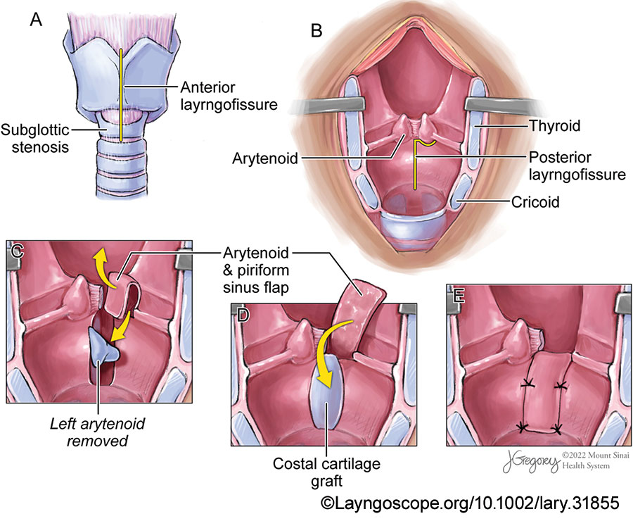
Figure 1: Expansion laryngoplasty steps: laryngofissure, arytenoidectomy, and reconstruction with costal cartilage graft and pyriform sinus flap. (A) A standard anterior laryngofissure is made in the midline to split the thyroid and cricoid cartilages. (B) The larynx is retracted open, and the cricoid is split posteriorly along with the posterior glottic stenosis in the interarytenoid space. (C) The arytenoidectomy is performed, but care is taken to preserve the arytenoid mucosa in what will become an anteriorly based piriform sinus flap. (D) The costal cartilage is inserted where the posterior cricoid split has been made and held in place by tension, then covered by the piriform sinus flap. (E) The pyriform sinus flap and cartilage are sutured in place to secure to reconstruction.

The surgery is divided into five main parts (Fig. 1):

  1. Rib cartilage harvest
  2. Laryngofissure and cricoid split
  3. Unilateral arytenoidectomy
  4. Pyriform sinus flap rotation and cartilage grafting
  5. Laryngeal stenting

A. Rib cartilage harvest: The procedure begins with a right-sided costal cartilage harvest. A costal cartilage graft of 3 cm × 1 cm × 0.5 cm is harvested. The design of the cartilage graft is similar to that described by Rutter and Cotton for pediatric stenosis. Briefly, a boat-shaped wedge of cartilage is harvested carefully to keep the periosteum on one surface.

B. Laryngofissure and cricoid split: A transverse incision is made, and cervical skin flaps are raised. The strap muscles are divided along the midline and retracted laterally. The tracheotomy site is evaluated and moved lower in the trachea if necessary. The perichondrium of the thyroid cartilage is then divided into the midline, and perichondrial flaps are preserved.

An oscillating saw is used to create the anterior laryngofissure through the thyroid and cricoid cartilages. The laryngofissure is completed using a #12 blade to incise the laryngeal mucosa without detaching either vocal fold from the thyroid cartilage (Fig. 1A). The posterior glottic scar and mucosa are divided sharply, followed by the splitting of the posterior cricoid ring. The lysis extends through the interarytenoid space and cuts the interarytenoid muscle (Fig. 1B). Subsequently, a complete split of the posterior cricoid ring is performed, and the cricoid ring is expanded to allow insertion of the costal cartilage graft.

C. Unilateral arytenoidectomy: Following the cricoid split and posterior commissure scar lysis, an arytenoidectomy is performed (Fig. 1C). Before surgery, the vocal fold with the least mobility or with the least favorable position is identified, and the arytenoid on that side is planned for resection. The mucosa overlying the arytenoid is carefully preserved by performing a submucosal arytenoidectomy. This mucosa becomes the pyriform sinus flap for coverage of the cricoid graft.

D. Pyriform sinus flap rotation and cartilage grafting: The pyriform sinus flap is created on the arytenoidectomy side by a releasing cut of the aryepiglottic fold laterally and a cut through the post-cricoid mucosa. This flap is cut to the level of the vocal fold. These cuts create a pedicle of arytenoid and medial pyriform sinus flap from the side of the arytenoidectomy. This mucosal flap is typically 2.5 cm in length and 1 cm in width. The flap is then rotated downward into the posterior lumen of the larynx to cover the costal cartilage graft (Fig. 1D). The mucosal flap must rotate to the bottom of the cricoid cartilage without tension. The superior height of the flap should not be lower than the opposite vocal fold.

The cartilage graft is placed into the field and fits into the posterior cricoid split defect. It is sutured in place with 4-0 Vicryl sutures, with the perichondrial side facing the airway lumen. The pyriform sinus flap is rotated down and secured with a Vicryl suture to cover the costal cartilage. Fibrin glue is then used (Fig. 1E).

Figure 2: Patient outcomes. Ten days after the original surgery, the patient was taken back to the operating room and the Montgomery laryngeal stent was removed. (A) The right pyriform sinus flap is well healed in the arytenoidectomy defect (arrow). (B) The left arytenoid and its position are preserved (arrow).

E. Laryngeal stenting: The Montgomery laryngeal stent is placed and secured. The anterior cricoid defect is augmented with costal cartilage if necessary. The laryngofissure is closed. The perichondrial flaps, strap muscles, and skin are closed. A Penrose drain is placed. A cuffed tracheotomy tube is then placed and secured. The stent can be removed 10 days after the initial surgery (Fig. 2). Decannulation can occur when the patient is capped without stridor.

RESULTS

From 2006 to 2021, the senior author (PW) performed EL on 11 patients, who all had combined glottic and subglottic stenosis. All the patients had high-grade glottis and infra-glottic stenosis with tracheotomy dependence. All have failed endoscopic interventions. Open surgery using cricoid expansion and/or arytenoidectomy was being considered. The average age of patients who underwent EL was 48.6 years (range 20-69 years). Of the 11 patients, five (45%) had diabetes mellitus, five (45%) had gastroesophageal reflux disease, and two (18%) had both.

Of the 11 patients who underwent EL, eight had undergone prior endoscopic or open procedures. These included endoscopic carbon dioxide laser excision of the scar with dilation and steroid injection, endoscopic partial arytenoidectomy, and thyroarytenoid myectomy with endoscopic transverse cordotomy. One of the patients had undergone prior laryngotracheoplasty. One patient had received a tracheal transplant but then had subsequent glottic and subglottic stenoses, thus preventing decannulation.

Figure 3: Patient example pre- and post-operatively. (A) Representative photo from in-office flexible fiberoptic laryngoscopy demonstrating complete glottic obstruction with phonation due to posterior glottic stenosis. (B) Representative photo from in-office flexible fiberoptic laryngoscopy one year after expansion laryngoplasty demonstrating a glottic airway of 3 mm posteriorly and (C) showing an open subglottic airway that has been expanded with the costal cartilage.

Of the 11 patients, eight (73%) were decannulated. Figure 3 is a pre- and postsurgical endoscopic view demonstrating complete pre-operative stenosis and the post-operative airway lumen from above and below. The remaining three patients could not be decannulated because of additional tracheal stenosis. The average time to decannulation was 4.6 months post-operatively (range one-13 months). Six patients required additional procedures, including endoscopic dilation and carbon dioxide laser trimming of the pyriform sinus flap after their EL. Only one patient reported post-operative dysphagia. This patient complained of aspiration with liquids 10 years after the initial surgery, which may be due to age-related swallowing dysfunction. She was treated with injection laryngoplasty into the prior arytenoid defect to build up the height of the posterior glottis. All the patients exhibited adequate voice with vibration sources from either the false vocal folds or the pyriform sinus flap. 

Filed Under: Head and Neck, How I Do It, Practice Focus Tagged With: EL, Expansion laryngoplasty

Leave a Reply Cancel reply

Your email address will not be published. Required fields are marked *

The Triological SocietyENTtoday is a publication of The Triological Society.

Polls

Would you choose a concierge physician as your PCP?

View Results

Loading ... Loading ...
  • Polls Archive

Top Articles for Residents

  • Applications Open for Resident Members of ENTtoday Edit Board
  • How To Provide Helpful Feedback To Residents
  • Call for Resident Bowl Questions
  • New Standardized Otolaryngology Curriculum Launching July 1 Should Be Valuable Resource For Physicians Around The World
  • Do Training Programs Give Otolaryngology Residents the Necessary Tools to Do Productive Research?
  • Popular this Week
  • Most Popular
  • Most Recent
    • The Dramatic Rise in Tongue Tie and Lip Tie Treatment

    • Rating Laryngopharyngeal Reflux Severity: How Do Two Common Instruments Compare?

    • Keeping Watch for Skin Cancers on the Head and Neck

    • Is Middle Ear Pressure Affected by Continuous Positive Airway Pressure Use?

    • Excitement Around Gene Therapy for Hearing Restoration

    • The Dramatic Rise in Tongue Tie and Lip Tie Treatment

    • Rating Laryngopharyngeal Reflux Severity: How Do Two Common Instruments Compare?

    • Is Middle Ear Pressure Affected by Continuous Positive Airway Pressure Use?

    • Otolaryngologists Are Still Debating the Effectiveness of Tongue Tie Treatment

    • Complications for When Physicians Change a Maiden Name

    • Reflections from a Past President of the Triological Society
    • ENT Surgeons Explore the Benefits and Challenges of AI-Powered Scribes: Revolutionizing Documentation in Healthcare
    • How To: Open Expansion Laryngoplasty for Combined Glottic and Subglottic Stenosis
    • How to: Pediatric Endoscopic Transsphenoidal Skull Base Approach and Closure: Technique and Technical Tips
    • TXA: Potentially Useful Adjunct in the Management of Anterior Epistaxis

Follow Us

  • Contact Us
  • About Us
  • Advertise
  • The Triological Society
  • The Laryngoscope
  • Laryngoscope Investigative Otolaryngology
  • Privacy Policy
  • Terms of Use
  • Cookies

Wiley

Copyright © 2025 by John Wiley & Sons, Inc. All rights reserved, including rights for text and data mining and training of artificial technologies or similar technologies. ISSN 1559-4939