INTRODUCTION
Large oronasal fistula (L-ONF) can severely affect patients’ feeding, speech, and mentality. However, for pediatric patients with L-ONF, surgical repair is difficult due to the lack of adjacent soft tissue and the low success rate of flap grafting (J Plast Surg Hand Surg. 2020;54:151–155). Thus, to improve the quality of life (QoL) of pediatric patients with L-ONF before a suitable time point for surgical repair, temporary prosthetic reconstruction of L-ONF with an obturator becomes a compromised choice.
Explore This Issue
June 2023To fabricate an obturator, the anatomic information of L-ONFs must be precisely obtained. However, the conventional impression-taking method may cause serious discomfort to patients due to irritation of the impression material to the sensitive nasal mucosa. To complicate matters, the impression material could simultaneously block the nasal and oral airways, resulting in dyspnea and even asphyxia (Dent J. 2014;2:142–154). These discomforts can aggravate noncompliance in children and increase challenges in obtaining an accurate impression.
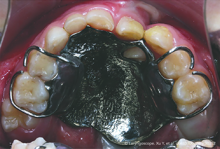
Figure 1. The oronasal fistula was restored with the 3D-printed hollow obturator.
To facilitate retention, obturators usually need to be crafted into a hollow structure to reduce weight. However, the conventional handmade process used by dental technicians is immensely complicated and time-consuming, which not only limits the accuracy of obturators but also increases the number of patient visits and the total treatment time.
Current innovations in computer-aided design (CAD) and computer-aided manufacturing are promising in breaking the abovementioned limitations. Intraoral scanning technologies are considered more comfortable, cost-effective, and precise than conventional impression techniques. In addition, additive manufacturing (3D printing) has been deemed a revolutionary technology in fabricating reconstructive prostheses with complex structures due to its advantages in customization, efficiency, and accuracy (Dent Mater. 2021;37:e314–e327).
METHOD
Patients
A 6-year-old girl with L-ONF after primary palatoplasty was enrolled in this study. QoL assessment was conducted before and six months after the obturator application. The 26-item version of the VeloPharyngeal Insufficiency Effects on Life Outcomes instrument was given to her parent for QoL assessment.
Digital Workflow
The digital workflow for the prosthetic reconstruction of L-ONFs with the 3D printed hollow obturator consists of four steps: 1) intraoral scanning, 2) CAD of the obturator, 3) additive manufacturing and postprocessing, and 4) obturator try-in.
Step 1: Intraoral Scanning. A confocal laser scanner was used for intraoral scanning. During scanning, the scanner tip was partially inserted into the tissue defect to capture the inner surface of the oronasal fistula.
Step 2: CAD Procedure. A hollow obturator was jointly designed by five CAD software programs. Briefly, the digital model generated by intraoral scanning was patched with connective bridges to obtain a closed surface. After smoothing the patched surface, a solid obturator was designed. The convex part of the obturator was allowed to partly enter the oronasal fistula to seal the tissue defect, and the concave surface of the obturator was designed to reconstruct the normal anatomic contour of the hard palate. After hollowing, the obturator was split into two parts for additive manufacturing: a hexagonal antirotation cover and a punched bottom (see Supplemental Video).
Step 3: Additive Manufacturing and Post-Processing. After data preprocessing, the cover and the bottom structure were separately printed using Ti-6Al-4V alloy powder by a laser powder bed fusion 3D printer. Thermal post-processing was conducted in a heat treatment furnace under argon protection. After support structure removal and sandblasting, the two printed parts were welded by a laser welding machine and then highly polished. Clasps were soldered onto the mesh area before acrylic resin packing.
Step 4: Obturator Try-In. The obturator was smoothly inserted into the patient’s fistula with an accurate fit and excellent sealing (Figure 1). The clasps of the obturator were slightly adjusted for better retention. Afterward, cone-beam computed tomography (CBCT) images were acquired for inspection.
RESULTS
Since the scanner tip did not contact the nasal or oral mucosa, the pediatric patient cooperated well and did not show any discomfort or behavioral resistance during intraoral scanning. The internal surface of the palatal fistula could be partly captured by the intraoral scanner, forming an open-edge hole in the center of the virtual model.
By CAD and additive manufacturing, the obturator was rapidly fabricated under numerical control. Compared to the conventional handmade technique, this digital workflow dramatically improved the manufacturing accuracy and simplified the fabrication process. Accordingly, the number of patient visits and the total treatment time were reduced. Due to the hollow design, the fabricated obturator weighed 4.1 g, which equals the weight of a dime.
The CBCT images indicated that the hollow obturator accurately fit the tissue defect. Both the nasal floor and oral roof were restored, separating the two cavities. After applying the obturator, the QoL of the patient was found to be improved, especially the eating and drinking dysfunctions listed in the domain of swallowing problems.