INTRODUCTION
With the introduction of sialendoscopy, a minimally invasive option now exists to treat patients with obstructive sialadenitis and other salivary ductal pathologies (N Engl J Med. 1999;341:1242-1243). Advances in technology have enabled providers of this technique to offer increasingly diverse options for benign salivary disease, including the introduction of calculi basket retrieval, stenosis dilation, and intraductal lithotripsy (Laryngoscope. 2012;122:1306-1311). The application of this diagnostic and therapeutic procedure has been limited by the technical challenges related to cannulation, dilation, and insertion of scope into the salivary duct.
Explore This Issue
August 2021Many dilator options have been devised to ease the introduction of the sialendoscope, yet the learning curve for these techniques has remained arduous. One early strategy included advancing a rigid, center-drilled bougie over a guidewire for papillary dilation (Laryngoscope. 2006;116:842–844). Serial rigid dilation with salivary dilators or lacrimal probes has been the main approach for papillary dilation for many years. More recently, proposed techniques include the use of an epidural tube with the epidural sheath used as the guidewire or utilizing a modified angiocatheter tip attached to the end of the scope to facilitate entrance (Laryngoscope. 2018;128:1392–1394). However, these techniques continue to require additional learned skills and significant anesthesia assistance. The addition of general anesthesia or deep sedation exposes patients to added, perhaps unnecessary, risk and may limit the use of sialendoscopy to therapeutic purposes only (i.e., for patients who have clear pathology discovered on preoperative assessments). With this mentality, sialendoscopy loses its effectiveness as a diagnostic study.
Herein, we demonstrate a novel, simplistic, and rapid approach to cannulation, dilation, and sialendoscope insertion that can be completed in an outpatient setting with the assistance of viscous lidocaine gel (see the supporting video).
METHOD
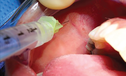
Figure 1. Application of local anesthesia via lidocaine injection in the parotid duct peri-papillary tissues.
Squires L, et al. Laryngoscope. 2021;131:E2432-E2435
Materials Needed: Surgeon’s choice of salivary endoscope (0.8, 1.1, 1.6 mm Erlangen, or 1.3 mm Marchal) with a camera head, a light source, and a video screen. A salivary access dilating catheter of choice from 4–0 Fr to 7–0 Fr is employed with assistance from a 0.4 mm or 0.01500 guidewire. Intraductal local anesthesia is employed with 2% viscous lidocaine gel, with or without peri-papillary topical cetacaine spray or injectional lidocaine. Saline irrigation (manual or powered) can be used to assist with the endoscopy. Cotton swabs (or intraoral retractors) and nontoothed forceps help facilitate access to the salivary ductal system.
Technique and Setup: Patients are seated in an awake, Fowler’s semi-supine, or an upright position. A self-retaining cheek retractor, dental block, or cotton swabs can be used to facilitate visualization of the papilla. When the patient is awake or under light sedation, there is often no need for retracting instrumentation, especially for submandibular duct papillae, as the patient can assist with visualization of the duct orifices. The first step begins with identification of the papilla followed by application of local anesthesia consisting of topical cetacaine spray, local 1% lidocaine injection with or without epinephrine (Figure 1), or 2% viscous lidocaine gel via cotton swabs. The authors have found this step to be optional even in the office setting. Once papillary anesthesia is obtained, a 0.4 mm or 0.01500 guidewire is then inserted into the papilla followed by the salivary access dilator via Seldinger technique to finish the papillary dilation. Debakey, Gerald, or other nontoothed forceps are useful at this step of the procedure to secure and advance the dilator to the desired level, generally at least 1 cm into duct. While the available salivary access dilating catheters range from 4–0 to 7–0 Fr, usually only one size is needed per case. The authors mainly utilize the 5–0 or 6–0 Fr sizes for the purpose of salivary papilla dilation and access, though the 4–0 Fr can be useful in cases with papillary stenosis and the 7–0 Fr catheter can help prepare the duct for passage of a larger endoscope, such as the Erlangen 1.6 mm.