• Home
  • Practice Focus
    • Facial Plastic/Reconstructive
    • Head and Neck
    • Laryngology
    • Otology/Neurotology
    • Pediatric
    • Rhinology
    • Sleep Medicine
    • How I Do It
    • TRIO Best Practices
  • Business of Medicine
    • Health Policy
    • Legal Matters
    • Practice Management
    • Tech Talk
    • AI
  • Literature Reviews
    • Facial Plastic/Reconstructive
    • Head and Neck
    • Laryngology
    • Otology/Neurotology
    • Pediatric
    • Rhinology
    • Sleep Medicine
  • Career
    • Medical Education
    • Professional Development
    • Resident Focus
  • ENT Perspectives
    • ENT Expressions
    • Everyday Ethics
    • From TRIO
    • The Great Debate
    • Letter From the Editor
    • Rx: Wellness
    • The Voice
    • Viewpoint
  • TRIO Resources
    • Triological Society
    • The Laryngoscope
    • Laryngoscope Investigative Otolaryngology
    • TRIO Combined Sections Meetings
    • COSM
    • Related Otolaryngology Events
  • Search

How To: Suspension Microesophagoscopy for Endoscopic Suture Closure of Pediatric Tracheoesophageal Fistula

by Evan J. Propst, MD, Jennifer M. Siu, MD, and Nikolaus E. Wolter, MD • July 11, 2024

  • Tweet
  • Click to email a link to a friend (Opens in new window) Email
Print-Friendly Version

INTRODUCTION

Tracheoesophageal fistula (TEF) without esophageal atresia, also termed H-type, N-type, Gross type E, or isolated fistula, can be congenital or acquired. Children with H-type TEF typically present with cough during feeding, cyanotic spells, and recurrent pneumonia. Endoscopic repair of H-type TEF via bronchoscopy has traditionally been limited to long, narrow (<2 mm), recurrent TEFs, which often require multiple procedures with interposed material (i.e., fibrin glue) and the occasional addition of ancillary procedures such as an esophageal balloon or a biosynthetic mesh plug (J Pediatr Surg. 2008;43:238-245. Curr Opin Otolaryngol Head Neck Surg. 2016;24:510-515). Wide (>2 mm), short TEFs are notoriously difficult to repair endoscopically and have traditionally required a thoracoscopic or open approach (Curr Opin Otolaryngol Head Neck Surg. 2016;24:510-515).

You Might Also Like

  • How To: H-Type Tracheoesophageal Fistula Cannulation for Rapid Intraoperative Localization
  • Early Otolaryngology Evaluation Advised for Neonates with Esophageal Atresia/ Tracheoesophageal Fistula
  • How To: Multilayered Closure of Oronasal and Oroantral Fistula Using Intranasal and Intraoral Flaps
  • How To: Oroantral Fistula Closure Using Double-Layered Flap
Explore This Issue
July 2024

We recently described suspending the esophagus with a laryngoscope to remove an intraluminal esophageal metal barbecue brush bristle in a child (Laryngoscope. 2021;131:E2066-E8). In that patient, the use of two hands under magnification with a wide field of view allowed the removal of a foreign body that would not have been accessible using a single instrument down a rigid esophagoscope. To our knowledge, suspension of the esophagus and the use of a microscope for endoscopic suture closure of a wide, short TEF have not previously been described. We report a patient for whom suspension microesophagoscopy was performed and describe how this approach allowed for improved management.

METHODS

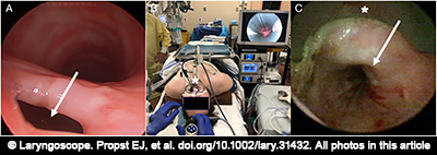
Figure 1. (A) Bronchoscopy demonstrating large, short tracheoesophageal fistula (TEF) (arrow). (B) Suspension esophagoscopy setup using Weerda distending laryngoscope (*). (C) Endoscopic view of esophagus in suspension with Weerda laryngoscope (*) showing exposure of TEF (arrow).

A 15-year-old male with prior Gross type C TEF repair, short stature, scoliosis, subaortic stenosis resection, ventricular septal defect repair, gastroesophageal reflux, and asthma was intubated due to parainfluenza A pneumonia and rhabdomyolysis. After five weeks of ventilation with a cuffed endotracheal tube, a tracheostomy was performed, with insertion of a size 6 Shiley DCT (cuffed with inner cannula). He was decannulated, but persistent aspiration pneumonia prompted flexible fiberoptic nasolaryngoscopy, which revealed a left vocal cord paralysis. Rigid bronchoscopy revealed an A-frame tracheal deformity and a large (4-mm diameter), short TEF just distal to it in the lower half of the trachea, likely in the location of the previous TEF repair (Fig. 1 ). This was a defect in the common party wall rather than a tract. The patient was referred to otolaryngology because he and his parents did not want to undergo open TEF repair, and it was felt that another long general anesthetic would not be well tolerated given his medical status. After discussing the very low likelihood that traditional transtracheal endoscopic methods (i.e., cautery and Tisseel) could close the large TEF, and that his rapid desaturations and narrow trachea would not allow suture closure from the tracheal side, the possibility of experimental suspension esophagoscopy and endoscopic suture repair was offered, and the family consented to the procedure.

Pages: 1 2 3 | Single Page

Filed Under: How I Do It, Laryngology, Practice Focus Tagged With: tracheoesophageal fistulaIssue: July 2024

You Might Also Like:

  • How To: H-Type Tracheoesophageal Fistula Cannulation for Rapid Intraoperative Localization
  • Early Otolaryngology Evaluation Advised for Neonates with Esophageal Atresia/ Tracheoesophageal Fistula
  • How To: Multilayered Closure of Oronasal and Oroantral Fistula Using Intranasal and Intraoral Flaps
  • How To: Oroantral Fistula Closure Using Double-Layered Flap

The Triological SocietyENTtoday is a publication of The Triological Society.

Polls

Would you choose a concierge physician as your PCP?

View Results

Loading ... Loading ...
  • Polls Archive

Top Articles for Residents

  • Applications Open for Resident Members of ENTtoday Edit Board
  • How To Provide Helpful Feedback To Residents
  • Call for Resident Bowl Questions
  • New Standardized Otolaryngology Curriculum Launching July 1 Should Be Valuable Resource For Physicians Around The World
  • Do Training Programs Give Otolaryngology Residents the Necessary Tools to Do Productive Research?
  • Popular this Week
  • Most Popular
  • Most Recent
    • A Journey Through Pay Inequity: A Physician’s Firsthand Account

    • The Dramatic Rise in Tongue Tie and Lip Tie Treatment

    • Otolaryngologists Are Still Debating the Effectiveness of Tongue Tie Treatment

    • Is Middle Ear Pressure Affected by Continuous Positive Airway Pressure Use?

    • Rating Laryngopharyngeal Reflux Severity: How Do Two Common Instruments Compare?

    • The Dramatic Rise in Tongue Tie and Lip Tie Treatment

    • Rating Laryngopharyngeal Reflux Severity: How Do Two Common Instruments Compare?

    • Is Middle Ear Pressure Affected by Continuous Positive Airway Pressure Use?

    • Otolaryngologists Are Still Debating the Effectiveness of Tongue Tie Treatment

    • Complications for When Physicians Change a Maiden Name

    • Excitement Around Gene Therapy for Hearing Restoration
    • “Small” Acts of Kindness
    • How To: Endoscopic Total Maxillectomy Without Facial Skin Incision
    • Science Communities Must Speak Out When Policies Threaten Health and Safety
    • Observation Most Cost-Effective in Addressing AECRS in Absence of Bacterial Infection

Follow Us

  • Contact Us
  • About Us
  • Advertise
  • The Triological Society
  • The Laryngoscope
  • Laryngoscope Investigative Otolaryngology
  • Privacy Policy
  • Terms of Use
  • Cookies

Wiley

Copyright © 2025 by John Wiley & Sons, Inc. All rights reserved, including rights for text and data mining and training of artificial technologies or similar technologies. ISSN 1559-4939