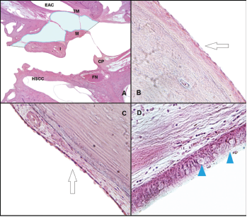
Histological analysis of the middle ear space. (A) Slide from a left ear, at the level of the pars flaccida, showing the space where the ciliated cells were counted at the epitympanum: lateral side of the malleus and incus, and medial side of the lateral wall. The light blue area represents the area where the ciliated cells were counted (H&E, 1 × magnification). (B) The arrow shows the flat epithelium located at the lateral side of the malleus (H&E, 10 × magnification). (C) The arrow shows the flat epithelium located at the medial side of the lateral wall at the epitympanum (H&E, 10 × magnification). (D) Histological view of the respiratory-like epithelium that covers the lateral wall of the protympanum toward the Eustachian tube. The arrowheads show the goblet cells located at this space (H&E, 40 × magnification). CP = cochleariform process; EAC = external auditory canal; FN = facial nerve; H&E = hematoxylin and eosin; HSCC = horizontal semicircular canal; I = incus; M = malleus; TM = tympanic membrane.
Credit: © 2018 The American Laryngological, Rhinological and Otological Society, Inc.