• Home
  • Practice Focus
    • Facial Plastic/Reconstructive
    • Head and Neck
    • Laryngology
    • Otology/Neurotology
    • Pediatric
    • Rhinology
    • Sleep Medicine
    • How I Do It
    • TRIO Best Practices
  • Business of Medicine
    • Health Policy
    • Legal Matters
    • Practice Management
    • Tech Talk
    • AI
  • Literature Reviews
    • Facial Plastic/Reconstructive
    • Head and Neck
    • Laryngology
    • Otology/Neurotology
    • Pediatric
    • Rhinology
    • Sleep Medicine
  • Career
    • Medical Education
    • Professional Development
    • Resident Focus
  • ENT Perspectives
    • ENT Expressions
    • Everyday Ethics
    • From TRIO
    • The Great Debate
    • Letter From the Editor
    • Rx: Wellness
    • The Voice
    • Viewpoint
  • TRIO Resources
    • Triological Society
    • The Laryngoscope
    • Laryngoscope Investigative Otolaryngology
    • TRIO Combined Sections Meetings
    • COSM
    • Related Otolaryngology Events
  • Search

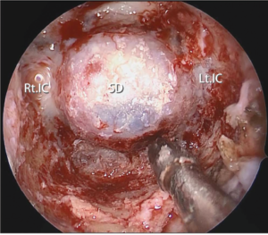
Fig. 1. Final endoscopic view after resection of the anterior wall of the sphenoid sinus including the PVC to the medial edge of the Vidian canal. Note the inferolateral part of the anterior sphenoidotomy is maximized, expanding the working corridor for surgical instruments. SD = sellar dura; Lt. IC = left internal carotid artery; Rt. IC = right internal carotid artery.
May 18, 2021

Print-Friendly Version

Fig. 1. Final endoscopic view after resection of the anterior wall of the sphenoid sinus including the PVC to the medial edge of the Vidian canal. Note the inferolateral part of the anterior sphenoidotomy is maximized, expanding the working corridor for surgical instruments. SD = sellar dura; Lt. IC = left internal carotid artery; Rt. IC = right internal carotid artery.

You Might Also Like

No related posts.

Fig. 1. Final endoscopic view after resection of the anterior wall of the sphenoid sinus including the PVC to the medial edge of the Vidian canal. Note the inferolateral part of the anterior sphenoidotomy is maximized, expanding the working corridor for surgical instruments. SD = sellar dura; Lt. IC = left internal carotid artery; Rt. IC = right internal carotid artery.

You Might Also Like:

The Triological SocietyENTtoday is a publication of The Triological Society.

Polls

Have you invented or patented something that betters the field of otolaryngology?

View Results

Loading ... Loading ...
  • Polls Archive

Top Articles for Residents

  • Applications Open for Resident Members of ENTtoday Edit Board
  • How To Provide Helpful Feedback To Residents
  • Call for Resident Bowl Questions
  • New Standardized Otolaryngology Curriculum Launching July 1 Should Be Valuable Resource For Physicians Around The World
  • Do Training Programs Give Otolaryngology Residents the Necessary Tools to Do Productive Research?
  • Popular this Week
  • Most Popular
  • Most Recent
    • The Dramatic Rise in Tongue Tie and Lip Tie Treatment

    • The Best Site for Pediatric TT Placement: OR or Office?

    • Rating Laryngopharyngeal Reflux Severity: How Do Two Common Instruments Compare?

    • Otolaryngologists Are Still Debating the Effectiveness of Tongue Tie Treatment

    • The Road Less Traveled—at Least by Otolaryngologists

    • The Dramatic Rise in Tongue Tie and Lip Tie Treatment

    • Rating Laryngopharyngeal Reflux Severity: How Do Two Common Instruments Compare?

    • Is Middle Ear Pressure Affected by Continuous Positive Airway Pressure Use?

    • Otolaryngologists Are Still Debating the Effectiveness of Tongue Tie Treatment

    • Complications for When Physicians Change a Maiden Name

    • Leaky Pipes—Time to Focus on Our Foundations
    • You Are Among Friends: The Value Of Being In A Group
    • How To: Full Endoscopic Procedures of Total Parotidectomy
    • How To: Does Intralesional Steroid Injection Effectively Mitigate Vocal Fold Scarring in a Rabbit Model?
    • What Is the Optimal Anticoagulation in HGNS Surgery in Patients with High-Risk Cardiac Comorbidities?

Follow Us

  • Contact Us
  • About Us
  • Advertise
  • The Triological Society
  • The Laryngoscope
  • Laryngoscope Investigative Otolaryngology
  • Privacy Policy
  • Terms of Use
  • Cookies

Wiley

Copyright © 2025 by John Wiley & Sons, Inc. All rights reserved, including rights for text and data mining and training of artificial technologies or similar technologies. ISSN 1559-4939

Fig. 1. Final endoscopic view after resection of the anterior wall of the sphenoid sinus including the PVC to the medial edge of the Vidian canal. Note the inferolateral part of the anterior sphenoidotomy is maximized, expanding the working corridor for surgical instruments. SD = sellar dura; Lt. IC = left internal carotid artery; Rt. IC = right internal carotid artery.