• Home
  • Practice Focus
    • Facial Plastic/Reconstructive
    • Head and Neck
    • Laryngology
    • Otology/Neurotology
    • Pediatric
    • Rhinology
    • Sleep Medicine
    • How I Do It
    • TRIO Best Practices
  • Business of Medicine
    • Health Policy
    • Legal Matters
    • Practice Management
    • Tech Talk
    • AI
  • Literature Reviews
    • Facial Plastic/Reconstructive
    • Head and Neck
    • Laryngology
    • Otology/Neurotology
    • Pediatric
    • Rhinology
    • Sleep Medicine
  • Career
    • Medical Education
    • Professional Development
    • Resident Focus
  • ENT Perspectives
    • ENT Expressions
    • Everyday Ethics
    • From TRIO
    • The Great Debate
    • Letter From the Editor
    • Rx: Wellness
    • The Voice
    • Viewpoint
  • TRIO Resources
    • Triological Society
    • The Laryngoscope
    • Laryngoscope Investigative Otolaryngology
    • TRIO Combined Sections Meetings
    • COSM
    • Related Otolaryngology Events
  • Search

How To: Transseptal Approach to the Maxillary Sinus and Pterygopalatine Fossa

by Camilo Reyes, MD, Brittany Gill, BS, and Lindsey Ryan, MD • August 16, 2022

  • Tweet
  • Click to email a link to a friend (Opens in new window) Email
Print-Friendly Version

Introduction

A range of open and endoscopic approaches to the maxillary sinus have been described and used for the treatment of both rhinological and skull base disease. Endoscopic approaches to the maxillary sinus have largely replaced open techniques, but in selected scenarios open approaches are still necessary (J Laryngol Otol. 2020;134:473–480).

You Might Also Like

  • How to: Nasoseptal Flap to Repair Large Maxillary Sinus Floor Defects
  • Integrating Preoperative Imaging, Intraoperative Findings in Maxillary Sinus Floor Infiltration Assessment in Patients with Maxillary Cancer
  • How to: Improving the Outcomes of Anterior Septal Perforations Repair with Combined Flaps
  • How To: Carolyn’s Window Approach to Unilateral Frontal Sinus Surgery
Explore This Issue
August 2022

While a wide variety of pathologies of the maxillary sinus, pterygopalatine, and infratemporal fossa can be addressed through an ipsilateral or an open approach, contralateral approaches are useful for lesions seated laterally, anteriorly, and inferiorly in the coronal plane. Expanded approaches to these anatomical areas include the Caldwell-Luc, mega-antrostomy, medial maxillectomy, Denker’s, prelacrimal Denker’s, and transseptal. A transseptal approach enables improved access to challenging anatomical locations, particularly pathology seated in the anterior and lateral regions (Am J Rhinol Allergy. 2009;23:426–432). However, access maybe be limited by the height of the nasal floor and there is an increased risk of septal perforation (J Laryngol Otol. 2020;134:473–480). Septal perforation symptoms can vary depending on the size and the location of the defect; however, they include epistaxis, nasal obstruction, crusting, nasal discharge, cacosmia, and intermittent whistling, and have a negative impact on a patient’s quality of life. They can be difficult to manage and are a risk associated with any septoplasty. This article describes a transseptal approach by elevating bilateral mucoperichondrial flaps and approaching a lesion from the contralateral side, which offers an expanded window to the maxillary sinus. Additionally, we also demonstrate our technique for septal reconstruction after this approach.

Method

A 40-year-old African American male presented to an outside facility with a six-month history of nasal obstruction. In-office biopsy reported a nasal polyp, and he was scheduled to undergo middle meatal antrostomy, septoplasty, and inferior turbinate reduction. Intraoperatively, he was found to have an inverted papilloma. His surgery was then aborted, and he was referred to Augusta University Medical Center for further management. The patient was scheduled for a transseptal approach to the maxillary sinus and pterygopalatine fossa with septoplasty.
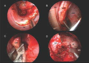
Figure 1. (A) Incision and elevation of “U”-shaped mucoperichondrial flap; the dotted line marks the area of incision. (B) Contralateral side mucoperichondrial flap sutured to the lateral nasal wall. (C) Removal of septal cartilage; the white arrow shows the transseptal window where the scope will pass. (D) Transseptal window showing exposure; the white dotted line marks the area where the “U”-shaped flap was raised. cNS = cartilaginous nasal septum; IP = inverted papilloma; IT = inferior turbinate; MPF = mucoperichondrial flap; MS = maxillary sinus; NF = nasal floor; NS = nasal septum; NV = nasal vestibule; UF = “U”-shaped flap.

Pages: 1 2 | Single Page

Filed Under: How I Do It, Practice Focus, Rhinology Tagged With: clinical best practices, clinical researchIssue: August 2022

You Might Also Like:

  • How to: Nasoseptal Flap to Repair Large Maxillary Sinus Floor Defects
  • Integrating Preoperative Imaging, Intraoperative Findings in Maxillary Sinus Floor Infiltration Assessment in Patients with Maxillary Cancer
  • How to: Improving the Outcomes of Anterior Septal Perforations Repair with Combined Flaps
  • How To: Carolyn’s Window Approach to Unilateral Frontal Sinus Surgery

The Triological SocietyENTtoday is a publication of The Triological Society.

Polls

Would you choose a concierge physician as your PCP?

View Results

Loading ... Loading ...
  • Polls Archive

Top Articles for Residents

  • Applications Open for Resident Members of ENTtoday Edit Board
  • How To Provide Helpful Feedback To Residents
  • Call for Resident Bowl Questions
  • New Standardized Otolaryngology Curriculum Launching July 1 Should Be Valuable Resource For Physicians Around The World
  • Do Training Programs Give Otolaryngology Residents the Necessary Tools to Do Productive Research?
  • Popular this Week
  • Most Popular
  • Most Recent
    • A Journey Through Pay Inequity: A Physician’s Firsthand Account

    • The Dramatic Rise in Tongue Tie and Lip Tie Treatment

    • Otolaryngologists Are Still Debating the Effectiveness of Tongue Tie Treatment

    • Is Middle Ear Pressure Affected by Continuous Positive Airway Pressure Use?

    • Rating Laryngopharyngeal Reflux Severity: How Do Two Common Instruments Compare?

    • The Dramatic Rise in Tongue Tie and Lip Tie Treatment

    • Rating Laryngopharyngeal Reflux Severity: How Do Two Common Instruments Compare?

    • Is Middle Ear Pressure Affected by Continuous Positive Airway Pressure Use?

    • Otolaryngologists Are Still Debating the Effectiveness of Tongue Tie Treatment

    • Complications for When Physicians Change a Maiden Name

    • Excitement Around Gene Therapy for Hearing Restoration
    • “Small” Acts of Kindness
    • How To: Endoscopic Total Maxillectomy Without Facial Skin Incision
    • Science Communities Must Speak Out When Policies Threaten Health and Safety
    • Observation Most Cost-Effective in Addressing AECRS in Absence of Bacterial Infection

Follow Us

  • Contact Us
  • About Us
  • Advertise
  • The Triological Society
  • The Laryngoscope
  • Laryngoscope Investigative Otolaryngology
  • Privacy Policy
  • Terms of Use
  • Cookies

Wiley

Copyright © 2025 by John Wiley & Sons, Inc. All rights reserved, including rights for text and data mining and training of artificial technologies or similar technologies. ISSN 1559-4939