• Home
  • Practice Focus
    • Facial Plastic/Reconstructive
    • Head and Neck
    • Laryngology
    • Otology/Neurotology
    • Pediatric
    • Rhinology
    • Sleep Medicine
    • How I Do It
    • TRIO Best Practices
  • Business of Medicine
    • Health Policy
    • Legal Matters
    • Practice Management
    • Tech Talk
    • AI
  • Literature Reviews
    • Facial Plastic/Reconstructive
    • Head and Neck
    • Laryngology
    • Otology/Neurotology
    • Pediatric
    • Rhinology
    • Sleep Medicine
  • Career
    • Medical Education
    • Professional Development
    • Resident Focus
  • ENT Perspectives
    • ENT Expressions
    • Everyday Ethics
    • From TRIO
    • The Great Debate
    • Letter From the Editor
    • Rx: Wellness
    • The Voice
    • Viewpoint
  • TRIO Resources
    • Triological Society
    • The Laryngoscope
    • Laryngoscope Investigative Otolaryngology
    • TRIO Combined Sections Meetings
    • COSM
    • Related Otolaryngology Events
  • Search

How To: Endoscopic Total Maxillectomy Without Facial Skin Incision

by Masato Nagaoka, MD, PhD, Kazuhiro Omura, MD, PhD, Taisuke Akutsu, MD, PhD, Haruyuki Hirayama, MD, Katsuhiro Ishida, MD, PhD, and Hiromi Kojima, MD, PhD • June 6, 2025

  • Tweet
  • Click to email a link to a friend (Opens in new window) Email
Print-Friendly Version

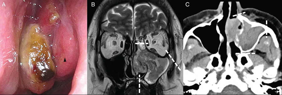
Figure 4: Pre-operative imaging findings of Case 3. (A) Left nasal findings. A tumor (asterisk) protruding from the maxillary sinus into the middle and common nasal passages was observed. The inferior turbinate (arrow) was identified. (B) Magnetic resonance imaging (T2-weighted image, coronal section). The tumor occupied the left maxillary sinus and partly protruded into a common nasal passage. The interior was heterogeneous, and there was no obvious destruction of the maxillary sinus bone. The medial orbital wall, hard palate, and zygomatic process were transected, as indicated by the white dotted line. (C) Computerized tomography (CT, horizontal section). Tumors were found in the maxillary sinus and partially connected to the nasal cavity. The nasal bone, zygomatic process, and pterygoid process were transected as the white dotted lines.

INTRODUCTION

Total maxillectomy is a surgical procedure involving amputation of the orbital floor, zygomatic bone, hard palate, and pterygoid process. It is typically performed in patients with cancer that has invaded the maxillary sinus. A common approach involves an external facial incision through a Weber–Ferguson skin incision; however, this method is associated with post-operative cosmetic complications. Furthermore, the conventional external incision approach is blind to the posterior and nasal sides of the maxillary sinus, which presents challenges in ensuring accurate margins. To compensate for these shortcomings, there are very few reports on endoscopically assisted surgery for advanced cancer of the maxillary sinus; however, there have been no reports of en bloc total maxillectomies in which a single tumor is resected without any facial skin incision. We developed a method for complete removal of the maxilla in cases of maxillary sinus cancer using an endoscopic technique that eliminates the need for facial skin incision. This technique ensures resection of the medial orbital wall and pterygoid process, which would otherwise be obscured by conventional methods.

You Might Also Like

  • How To: Full Endoscopic Procedures of Total Parotidectomy
  • Second Opinions: Facial Nerve Paralysis
  • Management of Malignant Tumors that Invade the Temporal Bone
  • How To: Meticulous Approach to Medial Palpebral Ligament in Lateral Rhinotomy: Avoiding Canthus Malposition
Explore This Issue
June 2025

In this study, three patients who underwent endoscopic maxillary surgery were examined. En bloc resection was successfully achieved in all three cases, with negative margins. The problem with endoscopic surgery is the difficulty in nasal manipulation; however, our co-authors have developed techniques to improve maneuverability. This study presents a pioneering report on an endoscopic total maxillectomy that allows for en bloc resection without a facial skin incision.

METHODS

Patients

Endoscopic total and subtotal maxillary sinus resection procedures were performed in three patients at Jikei University Hospital between April and October 2023. One patient with maxillary sinus cancer and two patients with oral cancer extending into the nasal cavity or maxillary sinus were included. Tumor location, stage, pathology, approach, surgical technique, and resection margins were comprehensively documented. The study was approved by the Ethics Committee (approval number: 34-102) and adhered to the ethical standards of the Human Experimentation Committee of Jikei University School of Medicine and the 1975 Declaration of Helsinki, as revised in 1983. We used an opt-out approach, and none of the patients refused consent.

All Instruments and Settings

We prepared a general nasal sinus endoscopic surgical instrument (Nagashima Medical Instruments, Tokyo, Japan). Endoscopic manipulation was performed using 4 mm rigid 0° and 70° endoscopes (Olympus Medical Systems, Tokyo, Japan). When cutting the mucous membranes of the nose, we used a microneedle electrode with a total length of 14 mm (Muranaka Medical Instruments, Osaka, Japan). When drilling the maxillary bone, a Midas Rex drill system (Medtronic, Minneapolis, USA) with a diamond bur was used to cut the nasal bone and hard palate. When cutting the maxillary bone, we used a high-speed Primado2 bone saw (NSK, Tokyo, Japan). When reconstructing bone, we used the AO Matrix MIDFACE Preformed Orbital Plate (Synthes, Oberdorf, Switzerland). The intra-operative setting was also shown (Fig. 1).

Figure 1: Intra-operative setting. Before resection: The otolaryngologist stands on the patient’s right side, with the endoscopic monitor on the head side. When operations can be performed simultaneously, the head and neck surgeon stands to the left of the patient to operate. The reconstructive surgeon also performed the surgical operation at the same time as the resection physician. Reconstruction: The reconstructive surgeon stands at the patient’s head side to operate.\

Surgical Technique

A three-dimensional model (LEXI Corporation, Tokyo, Japan) of the facial bone was created prior to surgery. Information on the extent of resection was shared between the head and neck surgery and plastic surgery teams. Considering the tumor location, a surgical passage through the nasal septum was created from the contralateral nasal cavity. An additional surgical passage through the anterior wall of the maxillary sinus was created if the anterior wall was not invaded. For lesions occupying the maxillary sinus, a transconjunctival approach was used with an eyelid conjunctival incision.

The contralateral sinus was opened during endoscopic manipulation. A nasal septal mucosal flap was created and preserved. The anterior wall of the sphenoid sinus was opened through the transnasal septum. The Draf III was performed, and the frontal sinuses were opened on both sides. The nasal septal cartilage was removed to widen the working space inside the nasal cavity on the lesion side. The pyriform aperture was exposed, and the nasolacrimal duct was dissected (Fig. 2A). The periorbita was exposed so as not to touch the tumor protruding into the nasal cavity. To reduce the operative time, endoscopic manipulation and oral incision were performed simultaneously. The anterior wall of the left maxilla was exposed, and the bone was exposed to the maxillary zygomatic ridge. Intraorally, a mucosal incision was made in the hard palate mucosa and around the pterygoid process, and the mandibular muscle process was removed. An eyelid conjunctival incision was made to reveal the orbital rim and expose the anterior wall of the maxillary sinus. The orbital rim and pyriform aperture were identified, and the nasal bone was cut endoscopically (Fig. 2B). The bone of the orbital floor was exposed, and the zygomatic process was cut with a bone saw with endoscopic assistance (Fig. 2C). The bone was exposed after a mucosal incision was made at the base of the nasal cavity. The median part of the hard palate was severed endoscopically, and the borders between the soft and hard palates and the Eustachian tube were also separated (Fig. 2D). The Vidian nerve was identified at the pterygoid base, the sphenopalatine artery was cauterized, and the pterygoid base was shaved from the nasal side (Fig. 2E). Subsequently, the tumor gained mobility, the residual pterygoid muscle was resected, and the mass was removed en bloc. The orbital floor was replaced with a titanium plate, and the maxillary defect was reconstructed with a free fibula. Excess bone protruding into the nasal cavity was shaved and adjusted endoscopically. Finally, the exposed bone was covered with a nasal septal mucocutaneous valve (Fig. 2F).

Figure 2: Nasal endoscopic surgical findings. (A) Findings in the left nose after nasal septal cartilage removal and right endoscopic surgical manipulation. The nasolacrimal duct at the medial wall of the orbit was cut (asterisk). (B) The orbital rim and pyriform aperture were identified, and the nasal bone was cut. (C) The bone of the orbital floor was exposed and cut with a bone saw while avoiding intraorbital fat (asterisk). (D) The bone was exposed on the nasal floor. The hard palate was endoscopically shaved and cut (dotted line). (E) The sphenoid sinus was open (asterisk). The base of the pterygoid process was drilled (dotted line). (F) The exposed fibula in the nasal cavity was endoscopically covered with a nasal septal mucocutaneous valve (asterisk).

RESULTS

Regarding endoscopic manipulation, a maximal three-port approach was used in Cases 1 and 2 to perform subtotal maxillectomy (Fig. 3). Case 3 involved a lesion occupying the maxillary sinus (Fig. 4); the zygomatic process was cut via two ports and a transconjunctival approach. A gingival incision was also made in all cases. In all cases, the tumor protruding into the nasal cavity was identified endoscopically and removed as one lump without incision.

Figure 3: Pre-operative imaging findings of Case 1. (A) Pre-operative intraoral findings. Tumor (arrow) bulge centered on the hard palate. (B) Right nasal findings. It was filled with a tumor (asterisk). (C) Magnetic resonance imaging (MRI; contrast-enhanced T1-weighted image, horizontal section). An internally heterogeneous tumor (asterisk) with partial contrast was observed, destroying the hard palate. (D) MRI (contrast-enhanced T1-weighted image, coronal section). A tumor (asterisk) was found in the nasal cavity, destroying and invading the hard palate. The tumor had partly extended into the interior of the maxillary sinus.

Case 1 was histopathologically diagnosed as myoepithelial carcinoma, and Cases 2 and 3 as squamous cell carcinoma, with negative resection margins in all cases, and no additional treatment required. The immediate post-operative and two-month post-operative facial appearances in Case 3 are shown (Fig. 5). The facial appearance and intranasal and oral findings of patients in cases 1 and 2 after surgery are shown (Fig. 6). No facial skin incisions were present, and patients were satisfied with the post-operative cosmetic outcomes. In all cases, the nasal cavity was covered with a mucous membrane, and there was minimal crusting.   

Figure 5: Photographs of Case 3 immediately after surgery (A–C) and two months after surgery (D–F). (A) The patient underwent a subconjunctival eyelid incision. After surgery, mild eyelid hyperemia and internal hemorrhage were observed. (B) The defect was reconstructed using a skin island obtained simultaneously with the fibula. (C) The tumor was resected in one lump, together with a tumor protruding medially into the nasal cavity. (D) Facial findings two months after surgery show no skin incision on the face. The lower eyelid is slightly retracted. (E) Contralateral inferior nasal dorsum (asterisk); a skin island (arrow) can be seen in the nasal cavity. A nasal septal valve (arrowhead) is used, and the interior of the nose is covered with a mucous membrane. (F) 3D CT imaging. The orbital floor was reconstructed with a titanium plate, and the interior of the maxillary sinus with fibula.

Figure 6: Photographs of Case 1, eight months after surgery (A-C) and Case 2, seven months after surgery (D-F). (A) Facial findings eight months after surgery show no skin incision on the face. (B) The defect was reconstructed using a skin island obtained simultaneously with the fibula. Skin islands (asterisk) conform to the surrounding tissue. (C) Contralateral inferior nasal dorsum (asterisk). A nasal septal valve (arrowhead) is used, and the interior of the nose is covered with a mucous membrane. (D) Facial findings seven months after surgery show no skin incision on the face. (a) The defect was reconstructed using a skin island obtained simultaneously with the fibula. Skin islands (asterisks) replace the nasal floor. Nasal septum removed, only the upper part (arrowhead) remaining. A nasal septal valve (arrow) is used, and the interior of the nose is covered with a mucous membrane. (F) 3D CT imaging. The orbital floor was reconstructed with a titanium plate, and the interior of the maxillary sinus with fibula. Most of the hard palate and the right maxillary sinus defect have been replaced by fibula.

Filed Under: Head and Neck, How I Do It, Practice Focus Tagged With: Endoscopic Total MaxillectomyIssue: June 2025

You Might Also Like:

  • How To: Full Endoscopic Procedures of Total Parotidectomy
  • Second Opinions: Facial Nerve Paralysis
  • Management of Malignant Tumors that Invade the Temporal Bone
  • How To: Meticulous Approach to Medial Palpebral Ligament in Lateral Rhinotomy: Avoiding Canthus Malposition

Leave a Reply Cancel reply

Your email address will not be published. Required fields are marked *

The Triological SocietyENTtoday is a publication of The Triological Society.

Polls

Would you choose a concierge physician as your PCP?

View Results

Loading ... Loading ...
  • Polls Archive

Top Articles for Residents

  • Applications Open for Resident Members of ENTtoday Edit Board
  • How To Provide Helpful Feedback To Residents
  • Call for Resident Bowl Questions
  • New Standardized Otolaryngology Curriculum Launching July 1 Should Be Valuable Resource For Physicians Around The World
  • Do Training Programs Give Otolaryngology Residents the Necessary Tools to Do Productive Research?
  • Popular this Week
  • Most Popular
  • Most Recent
    • A Journey Through Pay Inequity: A Physician’s Firsthand Account

    • The Dramatic Rise in Tongue Tie and Lip Tie Treatment

    • Rating Laryngopharyngeal Reflux Severity: How Do Two Common Instruments Compare?

    • Otolaryngologists Are Still Debating the Effectiveness of Tongue Tie Treatment

    • Is Middle Ear Pressure Affected by Continuous Positive Airway Pressure Use?

    • The Dramatic Rise in Tongue Tie and Lip Tie Treatment

    • Rating Laryngopharyngeal Reflux Severity: How Do Two Common Instruments Compare?

    • Is Middle Ear Pressure Affected by Continuous Positive Airway Pressure Use?

    • Otolaryngologists Are Still Debating the Effectiveness of Tongue Tie Treatment

    • Complications for When Physicians Change a Maiden Name

    • Excitement Around Gene Therapy for Hearing Restoration
    • “Small” Acts of Kindness
    • How To: Endoscopic Total Maxillectomy Without Facial Skin Incision
    • Science Communities Must Speak Out When Policies Threaten Health and Safety
    • Observation Most Cost-Effective in Addressing AECRS in Absence of Bacterial Infection

Follow Us

  • Contact Us
  • About Us
  • Advertise
  • The Triological Society
  • The Laryngoscope
  • Laryngoscope Investigative Otolaryngology
  • Privacy Policy
  • Terms of Use
  • Cookies

Wiley

Copyright © 2025 by John Wiley & Sons, Inc. All rights reserved, including rights for text and data mining and training of artificial technologies or similar technologies. ISSN 1559-4939