Malignant tumors that invade the temporal bone are rare, and diagnosis and treatment are straightforward with few areas of controversy. Nevertheless, these tumors often pose a surgical challenge, especially when they invade adjacent structures such as the carotid artery and the dura. ENToday spoke with experts in managing these uncommon but potentially disfiguring tumors about the current state of the art.
Explore This Issue
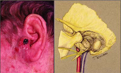
June 2007Tumors that can invade the temporal bone have three possible origins: the skin of the outer ear, the parotid gland, and the ear canal. Those that arise in the outer ear and parotid gland are the most common, whereas those arising in the ear canal are rare. Skin tumors present as a mass or ulcer in the outer ear, while parotid tumors present as a mass in the parotid gland. Tumors of the ear canal may present with signs that mimic chronic otitis media, such as hearing loss, purulence, or a canal mass. Other symptoms that can signal the presence of a tumor that invades the temporal bone are bleeding from the ear and facial paralysis or weakness.

“Any tumor that invades the temporal bone can produce purulent drainage, hearing loss, or affect the facial nerve,” said Paul W. Gidley, MD, Associate Professor at the Head and Neck Center at M. D. Anderson Cancer Center in Houston.
Diagnosis is made by physical examination of the head and neck, including thorough examination of the ear, parotid gland, neck, and cranial nerves and an evaluation of hearing. Imaging can assess the extent of the primary tumor and its relationship to adjacent structures, including the dura, brain tissue, facial nerve, and carotid artery. Imaging of the chest and the temporal bones and neck can assess the presence of metastases.
Often malignant tumors are misdiagnosed as an infection and treated with antibiotic drops. If a patient does not respond to treatment and has a mass in the ear canal, a biopsy should be obtained for diagnosis, said Barry Hirsch, MD, Professor of Otolaryngology at the University of Pittsburgh Eye and Ear Institute.
Preoperative Evaluation
Along with the overall health of the patient, the hearing status of the contralateral ear should be assessed. Staging should be done preoperatively by exam and imaging to provide some idea of the depth and invasion of the tumor. Both CT and magnetic resonance imaging provide complimentary information to assess the location and degree of tumor involvement. A CT scan can help determine whether bony erosion has occurred in the ear canal, whether the tumor has invaded the middle ear space, and if the tumor has spread to the temporomandibular joint. MRI can assess whether the parotid, upper neck lymph nodes, facial nerve, dura and brain, and soft tissues of the skull are involved, Dr. Hirsch explained.
Leave a Reply