INTRODUCTION
The goal of oncologic surgery is the complete resection of cancer with clear surgical margins. This is particularly important in head and neck cancer surgery, as the status of the surgical margin is the most significant pathologic risk factor (Head Neck Surg. 1978;1:107-111).
Explore This Issue
February 2023Margin assessment via frozen section analysis (FSA) is performed to ensure complete tumor resection with clear surgical margins while maximizing preservation of normal, uninvolved anatomical structures. A specimen-driven approach demands a higher level of intraoperative communication between surgeons and pathologists to convey the exact location of margin sampling. Given the complex, three-dimensional (3D) anatomy of head and neck oncologic specimens, surgeons and pathologists are often required to meet face to face to orient the specimen and discuss optimal margin sampling. This is not always feasible, however, because either the surgeon must leave the operating room (OR) or the pathologist must leave the gross room. FSA results are typically communicated back to the OR via telephone without any visual aid.
METHOD
3D Scanning
After an en bloc resection, the specimen was sent to the frozen section laboratory for specimen-driven margin analysis. A commercially available structured light desktop 3D scanner and accompanying software were used to capture and digitally reconstruct the 3D surface topography of fresh ex vivo surgical specimens. To optimize image quality, specimens were rinsed with water to remove blood products and patted dry, as blood products can interfere with the quality of image capture by the 3D scanner. Room lights were kept dim to optimize light exposure. A thin sheet of plastic was used to protect the scanner turntable. Specimens were serially imaged by the 3D scanner as the turntable platform completed a full revolution. The specimen was then flipped over to expose the opposite surface underneath, and the 3D capture process was repeated, creating two separate 3D data “point clouds.” Using EXScan, three-point cross-registration was performed to geometrically align each point cloud. The resulting meshwork was rendered into a watertight, photorealistic virtual 3D model of the en bloc resection specimen. The model was exported in 3MF file format into a computer-aided design (CAD) workspace.
Intraoperative Virtual 3D Specimen Mapping: The “CAD Margin”
The specimen was then handed off to the pathologist for standard-of-care FSA. Specimens were inked, sectioned, and processed into hematoxylin and eosin slides per standard protocol. Working side by side with the pathology prosector, a research assistant visualized the gross examination process and concurrently diagrammed the anatomic site of each sampled margin onto the 3D model using a paintbrush tool in the Meshmixer CAD workspace. Perpendicular and shave (en face) sections were graphically distinguished by color coding. Choice of perpendicular versus shave sectioning was at the pathologist’s and surgeon’s discretion. For our team, perpendicular margins are almost always used in the specimen-based margin approach, as they can measure distance from tumor to inked resection margin. Each section was labeled with a letter corresponding to the summary of sections listed within the final pathology report. The specimen prosector verified the accuracy of the 3D specimen map.
Surgical pathology fellows and/or attending pathologists were given access to the 3D specimen map during microscopy and determination of intraoperative margin status. In cases involving positive margins, the exact location of focal tumor presence was diagrammed onto the specimen map and highlighted with a unique color and label.
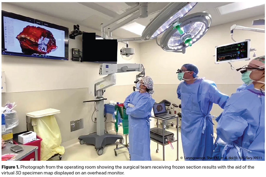
The CAD workspace, complete with 3D specimen map and frozen section diagnosis, was displayed on overhead OR monitors via remote videoconferencing software as a visual tool to facilitate surgeon–pathologist communication (Figure 1). Simultaneously, the surgical pathology fellows and/or attending pathologists verbalized the frozen section results. Surgeons utilized the 3D specimen maps to guide further resection and achieve negative final margin status when needed.
RESULTS
Intraoperative margin status was communicated with the aid of CAD specimen mapping in 12 cases. Median 3D image acquisition time was 7:18 (range 6:54–8:31). Operations consisted of six (50%) oral cavity composite resections, including two segmental mandibulectomies, two marginal mandibulectomies, and two without bony resection. Other operations included two (17%) transoral robotic surgeries (TORS), two (17%) partial glossectomies, one (8%) total laryngectomy, and one (8%) wide local cutaneous excision. Pathology was squamous cell carcinoma in all but one patient, who had a minor salivary gland (mammary analogue) secretory carcinoma of the buccal mucosa. Intraoperative margins were assessed by frozen section using a specimen-based approach in all cases.
Intraoperative specimen margin status was negative in four (33%) cases, and no further resection was required. The remaining eight cases involved close (<5 mm) (n = 3, 25%), focally positive (n = 3, 25%), or severely dysplastic (n = 2, 17%) margins initially seen on frozen section. 3D specimen maps successfully illustrated the anatomic site of each involved margin sampled from the en bloc resection specimen, thus providing visuospatial guidance for margin re-localization. Immediate re-resection was performed in five (42%) cases. Each case of re-resection resulted in a negative final margin status, although not all re-resection specimens contained malignancy.
In the two TORS cases, there were 1 mm and focally positive intraoperative margins. In case #9, the tumor was found to be extending through the superior constrictor on the main specimen with exposed external carotid artery in the defect; therefore further re-resection was not performed, resulting in an R1 final margin status. In the other TORS case, the superior constrictor was already resected in its entirety and freely mobile on the deep aspect of the main specimen, precluding further resection of the close (1 mm) margin. In case #8, frozen section showed an adequate 5-mm margin, which was found to be close (1 mm) on final pathology.