• Home
  • Practice Focus
    • Facial Plastic/Reconstructive
    • Head and Neck
    • Laryngology
    • Otology/Neurotology
    • Pediatric
    • Rhinology
    • Sleep Medicine
    • How I Do It
    • TRIO Best Practices
  • Business of Medicine
    • Health Policy
    • Legal Matters
    • Practice Management
    • Tech Talk
    • AI
  • Literature Reviews
    • Facial Plastic/Reconstructive
    • Head and Neck
    • Laryngology
    • Otology/Neurotology
    • Pediatric
    • Rhinology
    • Sleep Medicine
  • Career
    • Medical Education
    • Professional Development
    • Resident Focus
  • ENT Perspectives
    • ENT Expressions
    • Everyday Ethics
    • From TRIO
    • The Great Debate
    • Letter From the Editor
    • Rx: Wellness
    • The Voice
    • Viewpoint
  • TRIO Resources
    • Triological Society
    • The Laryngoscope
    • Laryngoscope Investigative Otolaryngology
    • TRIO Combined Sections Meetings
    • COSM
    • Related Otolaryngology Events
  • Search

How To: Modified Endoscopic Dacryocystorhinostomy Nasal Mucoperiosteal Flap Technique

by Tiechuan Cong, MD, Yuan Wu, MD, Ying Gao, MM, Quangui Wang, MM, and Yong Qin, MD • February 15, 2023

  • Tweet
  • Click to email a link to a friend (Opens in new window) Email
Print-Friendly Version

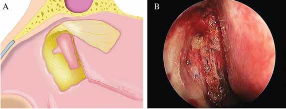
Figure 1. Making the bone window. (A) The bone around the maxillary line was drilled away and the medial wall of the lacrimal sac was exposed. Note the extended area of the bone window, facilitating the location of the anterior edge of the lacrimal sac. (B) Intraoperative view of the bone window and the exposed lacrimal sac.

Posteriorly hinged lacrimal mucoperiosteal flap: The medial wall of the sac was tented with a blunt Bowman’s lacrimal probe through the upper punctum and then incised vertically from the anterior-most margin with a crescent blade. Once the lacrimal sac was opened, the blade was inserted into the sac, and superior and inferior releasing incisions were made to create a posteriorly hinged lacrimal flap. The newly created lacrimal ostium was about 0.5 cm by 1 cm in size.

You Might Also Like

  • Is Post-­Operative Stenting Necessary in Endoscopic Dacryocystorhinostomy?
  • How to: Oroantral Fistula Closure Using Double-Layered Flap: Greater Palatine Artery Flap and Buccal Fat Pad
  • How To: Novel Endoscopic Technique to Repair Large Septal Perforation
  • Endoscopic Sandwich Technique for Moderate Nasal Septal Perforations
Explore This Issue
January 2023

Placement of two flaps: The lacrimal flap was reflected posteriorly and anastomosed with the mucoperiosteum overlying the bony uncinate process. Subsequently, the clubhead-shaped nasal flap was laid back from the olfactory cleft and repositioned over the bare bone.

Stent insertion (optional): A Crawford stent was inserted through upper and lower canaliculi and retrieved endoscopically. The stent was used in the condition of a severely narrowed lacrimal sac or stenotic canaliculus.

After the operation, small pieces of MeroGel pack were carefully placed into the marsupialized lacrimal sac, into the middle meatus, and over the lacrimal and nasal flaps to keep two flaps in apposition with the uncinate process and anterior edge of the opening of the lacrimal sac, respectively. The bare bone, about 0.5 cm by 1 cm, above the inferior turbinate was initially covered with Surgicel and then followed with MeroGel to facilitate secondary intension healing.

Postoperative Care

Postoperatively, three days of intravenous antibiotics, one week of antibiotic eye drops of tobramycin, and two months of intranasal corticosteroid were prescribed in all eDCRs. Lacrimal irrigation was not routinely performed. Patients were evaluated in terms of anatomical patency and symptom relief one week, three weeks, two months, and six months after surgery with nasal endoscopy. The stent remained in place for three months before being endoscopically removed.

RESULTS

Eight patients with nasolacrimal duct obstruction underwent clubhead-shaped flap eDCRs, all performed by the same surgeon, and were followed up at a mean period of 17.1 months. All patients had the symptom of epiphora except one, who was diagnosed with lacrimal sac mucocele and had only intermittent fullness in the medial canthal area. Surgical success was defined as the resolution of symptoms. Six patients with no stent insertion had relief of symptoms one week after the first nasal endoscopy and those with stent insertion three months after the stent removal. Two patients not living in Beijing were followed up only by telephone as a result of COVID-19. Both were persistently asymptomatic for 21 and 20 months after surgery, respectively. All of the patients had no recurrence and no marked complications.

Pages: 1 2 3 | Single Page

Filed Under: How I Do It, Practice Focus, Rhinology Tagged With: surgical managementIssue: January 2023

You Might Also Like:

  • Is Post-­Operative Stenting Necessary in Endoscopic Dacryocystorhinostomy?
  • How to: Oroantral Fistula Closure Using Double-Layered Flap: Greater Palatine Artery Flap and Buccal Fat Pad
  • How To: Novel Endoscopic Technique to Repair Large Septal Perforation
  • Endoscopic Sandwich Technique for Moderate Nasal Septal Perforations

The Triological SocietyENTtoday is a publication of The Triological Society.

Polls

Has experience as a patient influenced your professional development or demeanor?

View Results

Loading ... Loading ...
  • Polls Archive

Top Articles for Residents

  • A Resident’s View of AI in Otolaryngology
  • Call for Resident Bowl Questions
  • Resident Pearls: Pediatric Otolaryngologists Share Tips for Safer, Smarter Tonsillectomies
  • A Letter to My Younger Self: Making Deliberate Changes Can Help Improve the Sense of Belonging
  • ENTtoday Welcomes Resident Editorial Board Members
  • Popular this Week
  • Most Popular
  • Most Recent
    • A Case for Endoscopic Surgery: How Personal Experience Influenced Pursuit of a New Skill

    • Rewriting the Rules of Rhinosinusitis

    • Office Laryngoscopy Is Not Aerosol Generating When Evaluated by Optical Particle Sizer

    • Empty Nose Syndrome: Physiological, Psychological, or Perhaps a Little of Both?

    • Some Laryngopharyngeal Reflux Resists PPI Treatment

    • The Dramatic Rise in Tongue Tie and Lip Tie Treatment

    • Rating Laryngopharyngeal Reflux Severity: How Do Two Common Instruments Compare?

    • Is Middle Ear Pressure Affected by Continuous Positive Airway Pressure Use?

    • Otolaryngologists Are Still Debating the Effectiveness of Tongue Tie Treatment

    • Keeping Watch for Skin Cancers on the Head and Neck

    • Why So Loud? Rethinking the Volume of Our Everyday Experiences
    • How Audiologists and Researchers Are Shaping Military Hearing Health Practices
    • A Case for Endoscopic Surgery: How Personal Experience Influenced Pursuit of a New Skill
    • The Path to Department Chair: Arriving and Thriving
    • Rewriting the Rules of Rhinosinusitis

Follow Us

  • Contact Us
  • About Us
  • Advertise
  • The Triological Society
  • The Laryngoscope
  • Laryngoscope Investigative Otolaryngology
  • Privacy Policy
  • Terms of Use
  • Cookies

Wiley

Copyright © 2026 by John Wiley & Sons, Inc. All rights reserved, including rights for text and data mining and training of artificial technologies or similar technologies. ISSN 1559-4939