• Home
  • Practice Focus
    • Facial Plastic/Reconstructive
    • Head and Neck
    • Laryngology
    • Otology/Neurotology
    • Pediatric
    • Rhinology
    • Sleep Medicine
    • How I Do It
    • TRIO Best Practices
  • Business of Medicine
    • Health Policy
    • Legal Matters
    • Practice Management
    • Tech Talk
    • AI
  • Literature Reviews
    • Facial Plastic/Reconstructive
    • Head and Neck
    • Laryngology
    • Otology/Neurotology
    • Pediatric
    • Rhinology
    • Sleep Medicine
  • Career
    • Medical Education
    • Professional Development
    • Resident Focus
  • ENT Perspectives
    • ENT Expressions
    • Everyday Ethics
    • From TRIO
    • The Great Debate
    • Letter From the Editor
    • Rx: Wellness
    • The Voice
    • Viewpoint
  • TRIO Resources
    • Triological Society
    • The Laryngoscope
    • Laryngoscope Investigative Otolaryngology
    • TRIO Combined Sections Meetings
    • COSM
    • Related Otolaryngology Events
  • Search

How To: Superior Maximization of Sphenoidotomy with Olfaction Preservation in Endoscopic Endonasal Surgery

by Takeshi Hongo, MD, Yusuke Morinaga, MD, PhD, Hiroyoshi Kino, MD, PhD, Takuma Hara, MD, Takashi Kashiwagi, MD, PhD, Yasuhiro Tsunemi, MD, PhD, Makoto Akutsu, MD, PhD, Tsuguhisa Nakayama, MD, PhD, Shuho Tanaka, MD, PhD, and Hiroyoshi Akutsu, MD, PhD • September 3, 2025

  • Tweet
  • Click to email a link to a friend (Opens in new window) Email
Print-Friendly Version

INTRODUCTION

Endoscopic endonasal surgery (EES) for skull base lesions has advanced considerably. The transplanum transtuberculum approach is commonly used for anterior skull base lesions, where maximizing the anterior sphenoidotomy in the superior part is crucial for ensuring direct visualization and creating a wide working corridor. This procedure involves manipulation near the olfactory neuroepithelium; however, with a reported olfactory dysfunction risk. Although reports have detailed EES use for suprasellar lesions, methods for avoiding olfactory impairment remain unclear. A recent report revealed that EES for tuberculum sellae and planum sphenoidale meningiomas caused new post-operative anosmia in 21% of cases, warranting further investigations of the methods used to avoid olfactory impairment. The present report describes an EES technique that we devised for suprasellar lesions, which maximizes upper anterior sphenoidotomy while preserving the olfactory mucosa. We also evaluated the surgical outcomes, including a quantitative assessment of olfactory function.

You Might Also Like

  • How To: Opening the Palatovaginal Canal to Maximize Anterior Sphenoidotomy in Endoscopic Endonasal Surgery
  • How To: Vomer-Rostrum Mucosal Flap for Exposed Bone Coverage After Sphenoid Sinusotomy
  • How to: Pediatric Endoscopic Transsphenoidal Skull Base Approach and Closure: Technique and Technical Tips
  • A Closer Look at Endoscopic Ear Surgery
Explore This Issue
September 2025

METHODS

Study Design and Setting

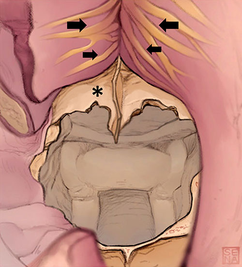
Figure 1. Schematic drawings showing the first step of superior maximization of anterior sphenoidotomy. After the nasal septal mucosa was carefully separated in a subperiosteal layer, the bony nasal septum was resected. The anterior wall of the sphenoid sinus and the vomer bone were exposed, and the ostium of the sphenoid sinus was identified. Olfactory filaments (arrows) are visible through the periosteum.

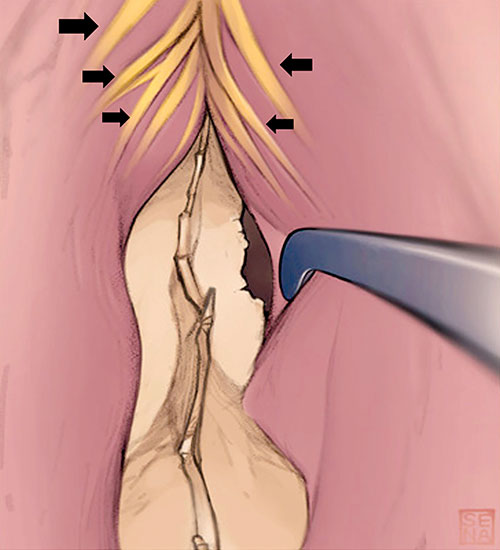
Figure 2. Schematic drawings showing the second step of superior maximization of anterior sphenoidotomy. The olfactory mucosa on the nasal septum was gently elevated, with olfactory filaments (arrows) visible through the periosteum. This procedure exposes the bony structure at the upper end of the anterior wall of the sphenoid sinus (asterisk), without damaging the olfactory mucosa.

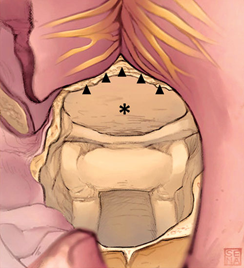
Figure 3. Schematic drawings showing the final step of superior maximization of anterior sphenoidotomy. The upper part of the anterior wall of the sphenoid sinus was completely removed up to the level of the skull base (arrowheads), with the olfactory mucosa fully preserved. After this procedure (maximizing the superior aspect of anterior sphenoidotomy), the planum sphenoidale (asterisk) was widely exposed and well visualized under a straight endoscope.

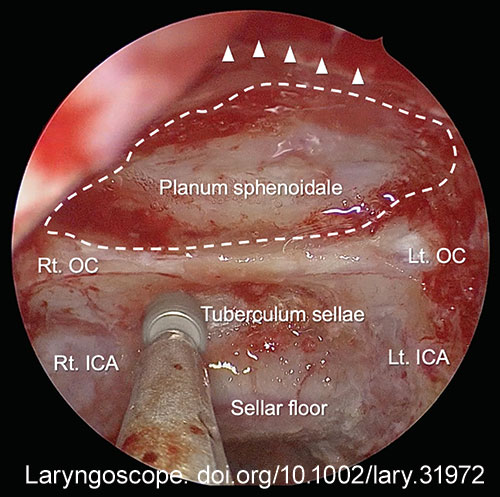
Figure 4. Endoscopic view after superior maximization of anterior sphenoidotomy in a representative case of tuberculum sellae meningioma. The anterior wall of the sphenoid sinus was removed to the level of the skull base (arrowheads). Panoramic view of the planum sphenoidale (dotted line) was obtained. Anatomical landmarks inside the sphenoid sinus are widely seen. Rt. OC = right optic canal; Lt. OC = left optic canal; Rt. ICA = right internal carotid artery; Lt. ICA = left internal carotid artery.

This retrospective observational study involved a prospectively collected database of adult patients who underwent initial EES for craniopharyngioma, tuberculum sellae, and planum sphenoidale meningiomas at the University of Tsukuba Hospital and Dokkyo Medical University Hospital between May 2015 and October 2022. Of the 56 patients, one who did not undergo the transplanum transtuberculum approach and 24 without pre- or post-operative quantitative olfactory assessments were excluded, leaving 31 patients. All patients underwent the presented surgical procedure to maximize upper anterior sphenoidotomy. Data on patient demographics and olfactory function were collected. The extent of resection was defined intra-operatively and radiographically as gross-total, near-total (95%–99%), subtotal (90%–94%), or partial (<90%). Written informed consent was obtained, and ethical approval was granted by the institutional review boards (R-52-8 J).

Evaluation Methods for Olfactory Function

Olfactory function was evaluated pre-operatively and at the six- and/or 12-month post-operative visits, prioritizing the 12-month evaluation when available. Olfactory function was assessed using a T&T olfactometer (Daiichi Pharmaceutical Co., Ltd., Tokyo, Japan), and severity was determined using odor recognition thresholds. This testing method has been verified to correlate with established olfactory evaluations, including the University of Pennsylvania Smell Identification Test. A threshold of ≤1.0 indicated normal function; 1.1–2.5, mild hyposmia; 2.6–4.0, moderate hyposmia; 4.1–5.5, severe hyposmia; and ≥5.6, anosmia. Olfactory function decline was defined as an increase of ≥1.0 in the threshold value.

Surgical Technique

The surgical technique of superior maximization of anterior sphenoidotomy with olfactory mucosa preservation is illustrated in Figures 1-3 and comprehensively through a representative case of tuberculum sellae meningioma in Figure 4.

Preparation

Spinal drainage was performed after general anesthesia induction when there was a high risk of post-operative cerebrospinal fluid leakage. The surgery was conducted by neurosurgeons and otolaryngologists using the bi-nostril three- or four-hand technique. Otolaryngologists performed nasal procedures, including sphenoid sinus opening and nasal reconstruction, while neurosurgeons performed the remaining procedures.

Exposure

The nasal septal mucosa was incised on one side using a Killian incision and detached under the cartilaginous periosteum on this side, while a horizontal posterior–superior mucosal incision was made on the other side. The perpendicular plates of the ethmoid bone and vomer, forming the bony nasal septum, were removed to expose the anterior wall of the sphenoid sinus (Fig. 1). The posterior ethmoid sinuses were accessed through the middle or superior nasal meatus via partial superior turbinectomy in the caudal one-third if necessary.

Wide Anterior Sphenoidotomy

The sphenoid sinus anterior wall was opened to a safe range with a rongeur, using the natural ostium as a landmark. The posterior wall mucosa of the sphenoid sinus was preserved for cranial base reconstruction. To achieve a broad aperture of the lower lateral segment of the anterior wall, the palatovaginal canals were opened, and additional removal was performed along the Vidian canal’s edge (the technical details are described in our previous report). Before removing the upper anterior wall, the nasal septal mucosa dissection was extended superiorly, exposing the olfactory mucosa in the upper portion of the nasal septum. Here, the olfactory filaments were visualized as white, thread-like structures on the undersurface of the septal mucosa, visible through the periosteum (Fig. 2). The olfactory mucosa of the nasal septum was carefully elevated from beneath the periosteum to the tegmen, avoiding olfactory nerve filament damage. This allowed the complete removal of the bony structures independent of the nasal septal mucosa. The perpendicular plate of the ethmoid bone and the anterior wall of the sphenoid sinus were removed until they joined the skull base (Fig. 3). This procedure rendered the tegmen of the nasal cavity continuous with the sphenoid sinus ceiling, providing panoramic exposure to the upper space within the sphenoid sinus. The sphenoid sinus septal walls were then sufficiently removed until vital anatomical landmarks were visible (Fig. 4).

Subsequent Procedures and Post-Operative Management

The bone covering the cellar floor and extending to the planum sphenoidale was widely resected. Following tumor resection, the dura was closed and covered with either a pedicled sphenoid sinus mucosal flap or a vascularized nasoseptal flap. The preserved olfactory mucosa was returned to its anatomical position, and absorbable dressings were placed within the olfactory cleft to prevent mucosal adhesion. An otolaryngologist routinely managed post-operative nasal care.

RESULTS

The median pre- and post-operative odor recognition thresholds were 1.6 and 2.0, respectively (p = 0.03). The olfactory function declined by more than one recognition threshold in nine patients (29%) post-operatively. Among these, post-operative olfactory dysfunction manifested as severe hyposmia in one patient, moderate hyposmia in two patients, mild hyposmia in five patients, and normal in one patient. Specifically, three patients (10%) experienced a decline in olfactory function to moderate or severe hyposmia; however, none of the patients developed anosmia. The mean age of patients with and without declining olfaction was 57 and 53 years (p = 0.53), and the percentages of meningiomas were 78% and 46% (p = 0.13), respectively. Additionally, there were no significant differences in the tumor diameter, extent of resection, or nasal procedures.

CONCLUSIONS

We presented a novel surgical method for superior maximization of anterior sphenoidotomy with preservation of the olfactory function in EES for suprasellar lesions. Our technique achieves maximal anterior sphenoidotomy with minimal negative impact on olfactory function and offers a viable option for surgeons concerned about the potential risks of olfactory impairment from wide sphenoidotomy. 

Filed Under: Articles, How I Do It, How I Do It, Practice Focus, Rhinology Tagged With: EES, Endoscopic endonasal surgery, skull base lesionsIssue: September 2025

You Might Also Like:

  • How To: Opening the Palatovaginal Canal to Maximize Anterior Sphenoidotomy in Endoscopic Endonasal Surgery
  • How To: Vomer-Rostrum Mucosal Flap for Exposed Bone Coverage After Sphenoid Sinusotomy
  • How to: Pediatric Endoscopic Transsphenoidal Skull Base Approach and Closure: Technique and Technical Tips
  • A Closer Look at Endoscopic Ear Surgery

Leave a Reply Cancel reply

Your email address will not be published. Required fields are marked *

The Triological SocietyENTtoday is a publication of The Triological Society.

Polls

Have you experienced an increase in in-office rhinology procedures in the last year?

View Results

Loading ... Loading ...
  • Polls Archive

Top Articles for Residents

  • ENTtoday Welcomes Resident Editorial Board Members
  • Applications Open for Resident Members of ENTtoday Edit Board
  • How To Provide Helpful Feedback To Residents
  • Call for Resident Bowl Questions
  • New Standardized Otolaryngology Curriculum Launching July 1 Should Be Valuable Resource For Physicians Around The World
  • Popular this Week
  • Most Popular
  • Most Recent
    • The Dramatic Rise in Tongue Tie and Lip Tie Treatment

    • Rating Laryngopharyngeal Reflux Severity: How Do Two Common Instruments Compare?

    • Keeping Watch for Skin Cancers on the Head and Neck

    • Physician, Know Thyself! Tips for Navigating Mid-Career Transitions in Otolaryngology

    • History of the Cochlear Implant

    • The Dramatic Rise in Tongue Tie and Lip Tie Treatment

    • Rating Laryngopharyngeal Reflux Severity: How Do Two Common Instruments Compare?

    • Is Middle Ear Pressure Affected by Continuous Positive Airway Pressure Use?

    • Otolaryngologists Are Still Debating the Effectiveness of Tongue Tie Treatment

    • Complications for When Physicians Change a Maiden Name

    • Questions on NIH Funding Leave ENT Researchers Pondering Next Steps and Leaving Everything Up in the Air
    • In-Office Rhinology Practices Continue to Grow
    • How Do We Define “Winning” in the OR?
    • A Letter to My Younger Self: Making Deliberate Changes Can Help Improve the Sense of Belonging
    • How To: Superior Maximization of Sphenoidotomy with Olfaction Preservation in Endoscopic Endonasal Surgery

Follow Us

  • Contact Us
  • About Us
  • Advertise
  • The Triological Society
  • The Laryngoscope
  • Laryngoscope Investigative Otolaryngology
  • Privacy Policy
  • Terms of Use
  • Cookies

Wiley

Copyright © 2025 by John Wiley & Sons, Inc. All rights reserved, including rights for text and data mining and training of artificial technologies or similar technologies. ISSN 1559-4939