• Home
  • Practice Focus
    • Facial Plastic/Reconstructive
    • Head and Neck
    • Laryngology
    • Otology/Neurotology
    • Pediatric
    • Rhinology
    • Sleep Medicine
    • How I Do It
    • TRIO Best Practices
  • Business of Medicine
    • Health Policy
    • Legal Matters
    • Practice Management
    • Tech Talk
    • AI
  • Literature Reviews
    • Facial Plastic/Reconstructive
    • Head and Neck
    • Laryngology
    • Otology/Neurotology
    • Pediatric
    • Rhinology
    • Sleep Medicine
  • Career
    • Medical Education
    • Professional Development
    • Resident Focus
  • ENT Perspectives
    • ENT Expressions
    • Everyday Ethics
    • From TRIO
    • The Great Debate
    • Letter From the Editor
    • Rx: Wellness
    • The Voice
    • Viewpoint
  • TRIO Resources
    • Triological Society
    • The Laryngoscope
    • Laryngoscope Investigative Otolaryngology
    • TRIO Combined Sections Meetings
    • COSM
    • Related Otolaryngology Events
  • Search

How To: Vomer-Rostrum Mucosal Flap for Exposed Bone Coverage After Sphenoid Sinusotomy

by Andrew J. Goates, MD, Garret Choby, MD, Quangui Wang, MM, and Carlos D. Pinheiro-Neto, MD • February 14, 2023

  • Tweet
  • Email
Print-Friendly Version

INTRODUCTION 

The differential diagnosis for sphenoid sinus opacification is broad and includes benign, malignant, and infectious etiologies. Patients may present with an atypical headache, retroorbital eye pain, visual disturbances, or sinonasal complaints, or it may be asymptomatic and incidentally noted on imaging for another indication. Generally, persistent sphenoid sinus opacification is evaluated and treated surgically with a sphenoid sinusotomy and biopsy as indicated. 

You Might Also Like

  • How To: Inferior Meatus Mucosal Flap for Septal Reconstruction and Resurfacing After Nasoseptal Flap Harvest
  • How to: Nasoseptal Flap to Repair Large Maxillary Sinus Floor Defects
  • How to: Oroantral Fistula Closure Using Double-Layered Flap: Greater Palatine Artery Flap and Buccal Fat Pad
  • Upper Lateral Cartilage Mucosal Flap Enables the Successful Closure of Larger Septal Perforations
Explore This Issue
February 2023

Sphenoid sinus opacification after operative intervention can reoccur due to scarring or bone remodeling and is reported to happen in 10% of cases, a rate that may be even more pronounced in the setting of mucoceles and fungus balls (Int J Surg Case Rep. 2021;79:18-23). The bone remodeling phenomenon has been described in the frontal sinus with restenosis following endoscopic modified Lothrop procedures (Int Forum Allergy Rhinol. 2019;9:1387-1394). Recent studies have found that placement of vascularized flaps or mucosal grafts resulted in reduced rates of stenosis and neo-ossification (Int Forum Allergy Rhinol. 2019;9:1387-1394).

METHOD

Operative Set-up

Patients are prepped and draped in the typical fashion for endoscopic surgery with exposure of the face. Computed tomography image guidance is registered using skin surface matching and is calibrated to within 1 to 2 mm of accuracy. The entire procedure is performed using a 0° endoscope.

Posterior Ethmoidectomy and Sphenoid Sinusotomy

Typically, the dissection is started on the side of the pathology. The inferior and middle turbinates are gently lateralized to improve the space within the nasal cavity and open access to the sphenoethmoidal recess. Any synechiae from prior surgery is carefully divided and the superior turbinate is identified. The location of the sphenoid sinus is confirmed using the computer navigation system, which can be helpful in the setting of severe scarring and bone remodeling. A retrograde posterior ethmoidectomy is performed using Kerrison rongeurs and microdebrider to expose the anterior wall of the sphenoid. Thru-cutting forceps are used to resect the inferior one-third of the superior turbinate. The region of the ostium of the sphenoid sinus is entered and dilated with the Cottle. A Kerrison rongeur is used to enlarge the sphenoidotomy with removal of the anterior wall of the sphenoid sinus superiorly toward the planum and laterally toward the orbit. At this point, purulence, fungal elements, inspissated secretions, or other pathology may be encountered and require culture, biopsy, irrigation, or suctioning.

Vomer-Rostral Flap Design and Elevation

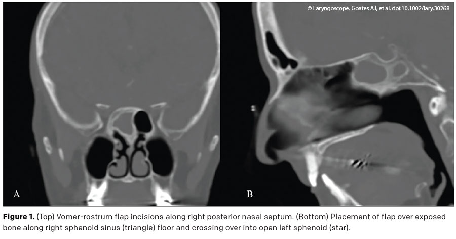
The mucosa over the sphenoid rostrum and nasal septum is elevated to expose the sphenoid rostrum bone. A rescue flap incision is performed with a needle-tip Bovie set at 10 W. This incision is started at the sphenoid sinus ostium and progresses anteriorly along the septal mucosa for about 1 to 2 cm and parallel to the palate. Then, the incision is turned inferiorly at a 90° angle toward the nasal floor, taken to the level of the superior border of the choana. An inferior mucosal incision is performed from the superior arch of the choana and carried anteriorly to join with the anterior incision. Using a Cottle dissector, the mucosal flap from the sphenoid rostrum is elevated with preservation of the vascular pedicle. The mucosal flap along the sphenoid rostrum containing the posterior septal artery is mobilized inferiorly, exposing the underlying bone (see Figure 1).

Pages: 1 2 | Single Page

Filed Under: How I Do It, Practice Focus, Rhinology Tagged With: rhinologyIssue: February 2023

You Might Also Like:

  • How To: Inferior Meatus Mucosal Flap for Septal Reconstruction and Resurfacing After Nasoseptal Flap Harvest
  • How to: Nasoseptal Flap to Repair Large Maxillary Sinus Floor Defects
  • How to: Oroantral Fistula Closure Using Double-Layered Flap: Greater Palatine Artery Flap and Buccal Fat Pad
  • Upper Lateral Cartilage Mucosal Flap Enables the Successful Closure of Larger Septal Perforations

The Triological SocietyENTtoday is a publication of The Triological Society.

Polls

Have you invented or patented something that betters the field of otolaryngology?

View Results

Loading ... Loading ...
  • Polls Archive

Top Articles for Residents

  • Applications Open for Resident Members of ENTtoday Edit Board
  • How To Provide Helpful Feedback To Residents
  • Call for Resident Bowl Questions
  • New Standardized Otolaryngology Curriculum Launching July 1 Should Be Valuable Resource For Physicians Around The World
  • Do Training Programs Give Otolaryngology Residents the Necessary Tools to Do Productive Research?
  • Popular this Week
  • Most Popular
  • Most Recent
    • The Dramatic Rise in Tongue Tie and Lip Tie Treatment

    • Otolaryngologists Are Still Debating the Effectiveness of Tongue Tie Treatment

    • The Best Site for Pediatric TT Placement: OR or Office?

    • Rating Laryngopharyngeal Reflux Severity: How Do Two Common Instruments Compare?

    • Keeping Watch for Skin Cancers on the Head and Neck

    • The Dramatic Rise in Tongue Tie and Lip Tie Treatment

    • Rating Laryngopharyngeal Reflux Severity: How Do Two Common Instruments Compare?

    • Is Middle Ear Pressure Affected by Continuous Positive Airway Pressure Use?

    • Otolaryngologists Are Still Debating the Effectiveness of Tongue Tie Treatment

    • Complications for When Physicians Change a Maiden Name

    • Leaky Pipes—Time to Focus on Our Foundations
    • You Are Among Friends: The Value Of Being In A Group
    • How To: Full Endoscopic Procedures of Total Parotidectomy
    • How To: Does Intralesional Steroid Injection Effectively Mitigate Vocal Fold Scarring in a Rabbit Model?
    • What Is the Optimal Anticoagulation in HGNS Surgery in Patients with High-Risk Cardiac Comorbidities?

Follow Us

  • Contact Us
  • About Us
  • Advertise
  • The Triological Society
  • The Laryngoscope
  • Laryngoscope Investigative Otolaryngology
  • Privacy Policy
  • Terms of Use
  • Cookies

Wiley

Copyright © 2025 by John Wiley & Sons, Inc. All rights reserved, including rights for text and data mining and training of artificial technologies or similar technologies. ISSN 1559-4939