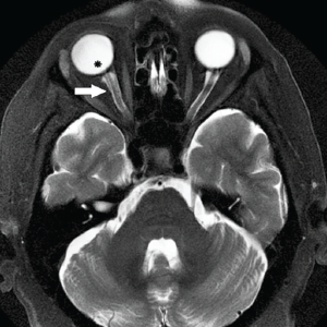
Axial, T2-weighted MRI demonstrating optic nerve sheath dilation (white arrow) and flattening of the posterior aspect of the right globe (asterisk). April 8, 2018 Print-Friendly Version You Might Also Like No related posts. Axial, T2-weighted MRI demonstrating optic nerve sheath dilation (white arrow) and flattening of the posterior aspect of the right globe (asterisk).