• Home
  • Practice Focus
    • Facial Plastic/Reconstructive
    • Head and Neck
    • Laryngology
    • Otology/Neurotology
    • Pediatric
    • Rhinology
    • Sleep Medicine
    • How I Do It
    • TRIO Best Practices
  • Business of Medicine
    • Health Policy
    • Legal Matters
    • Practice Management
    • Tech Talk
    • AI
  • Literature Reviews
    • Facial Plastic/Reconstructive
    • Head and Neck
    • Laryngology
    • Otology/Neurotology
    • Pediatric
    • Rhinology
    • Sleep Medicine
  • Career
    • Medical Education
    • Professional Development
    • Resident Focus
  • ENT Perspectives
    • ENT Expressions
    • Everyday Ethics
    • From TRIO
    • The Great Debate
    • Letter From the Editor
    • Rx: Wellness
    • The Voice
    • Viewpoint
  • TRIO Resources
    • Triological Society
    • The Laryngoscope
    • Laryngoscope Investigative Otolaryngology
    • TRIO Combined Sections Meetings
    • COSM
    • Related Otolaryngology Events
  • Search

How To: Full Endoscopic Procedures of Total Parotidectomy

by Jia-Lu He, MM, Xue-Er Zhou, MM, Chang Cao, MM, Su Chen, PhD, Fan Yang, PhD, Yong-Tao Dong, BS, Yan Sun, BS, and Gui-Quan Zhu, MD • May 6, 2025

  • Tweet
  • Email
Print-Friendly Version

INTRODUCTION

Parotid tumors (PTs) account for approximately 64% to 80% of salivary gland tumors, with the majority being benign and located in the superficial lobe. Generally, conservative surgical resection is the primary treatment for benign PTs, while total parotidectomy is recommended for malignant parotid lesions to obtain safe histological margins. Following the total parotidectomy, the sternocleidomastoid muscle flap (SCMF) can be used to prevent Frey’s syndrome and partially restore facial contour defects.

You Might Also Like

  • Second Opinions: Facial Nerve Paralysis
  • Salivary Gland Malignancies: Diagnosis and Treatment of a Rare and Challenging Cancer
  • Benefits, Drawbacks of Free Abdominal Fat Transfer for Parotidectomy Defect Reconstruction
  • How To: Endoscope-Assisted Transoral Procedure of Accessory Parotid Gland Tumor Resection
Explore This Issue
May 2025

Conventional total parotidectomy is typically conducted through a Blair incision, which can leave a visible facial scar, impacting the cosmetic satisfaction and quality of life of some patients. Recently, endoscopic surgery has been increasingly applied to head and neck surgery to achieve minimally invasive effects and improved aesthetic outcomes. There are few reports on the full endoscopic technique of total parotidectomy followed by SCMF transplantation to our knowledge, however.

We aim to describe the surgical procedures of full endoscopic total parotidectomy (FETP) and assess patient outcomes to evaluate the feasibility and efficacy of this novel approach.

MATERIALS AND METHODS

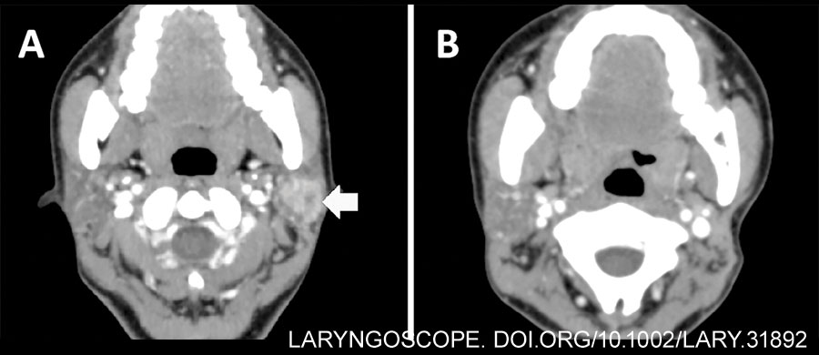
A case of parotid malignancy managed with FETP and SCMF transplantation at our department was retrospectively reviewed. Pre-operative contrast-enhanced CT revealed a 2×2 cm-sized mass with heterogeneous enhancement in the left superficial parotid gland (Fig. 1A). Preparation of hair removal involved a 2-3 cm area in the temporal region above the hairline and the postauricular region behind the hairline.

Figure 1: Pre-operative and post-operative contrast-enhanced CT. (A) Pre-operative contrast-enhanced CT of left parotid mass (horizontal arrow). (B) Post-operative contrast-enhanced CT images with no sign of recurrence (two years after surgery).

The operation was conducted according to the Declaration of Helsinki (as revised in 2013) and was approved by the Ethics Committee of West China Hospital of Stomatology, Sichuan University (Approval No. WCHSIRB-D-2020-311-R1). The patient was fully informed about the benefits and drawbacks of this procedure, as well as risks of conversion to conventional open surgery.

Surgical Technique

Following general endotracheal anesthesia and routine disinfection, the terminals of the neuromonitor were placed into the subcutaneous tissue of the manubriosternal region, orbicularis oculi, and orbicularis oris, respectively, for intra-operative facial nerve monitoring (Fig. 2A). To improve the post-operative aesthetic results, an approximately 5-cm incision was designed along the postauricular hairline region, with the reflection of neoplasm marked (Fig. 2B). Noticeably, a 1-cm temporal incision and cavity were designed to accommodate the suction device or other endoscopic instruments, facilitating cavity separation, blood suction, and tissue lifting. We referred to these designs as the “plus” incision and cavity in FETP surgery. The electrocautery, sterile cotton swab, and skin retractor were used when incising the skin and subcutaneous tissue (Fig. 2C). Following the creation of the main working cavity, a surgical retractor system was used to lift the skin flap stably and maintain a wide operative cavity (Fig. 2D). Then we expanded the main working cavity using a 5-mm laparoscope with 0° viewing angles and miniature instruments. Manipulation at this step may be obstructed by the cartilage of the external auditory canal. The “plus” cavity can then be established with blunt dissection to proceed with the separation of the superficial musculoaponeurotic system and achieve a distinct anatomical layer (Fig. 2E). Incise the connected fascia under endoscopic view until two cavities are fully merged. Additionally, stitches can be used to pull the earlobe area, achieving a larger operative cavity (Fig. 2F). To reduce post-operative numbness in the auricular skin, careful protection of the great auricular nerve (GAN) is crucial during the extension of the surgical tunnel (Fig. 3A).

Figure 2: Pre-operative preparation and the creation of surgical cavity under direct view. (A) The placement of terminals of neuromonitor. (B) The design of incisions and working cavity. (C) Incise the skin under direct view. (D) The use of surgical retractor systems. (E) Fusion of two surgical cavities. (F) Pull the earlobe to enlarge the surgical cavity.

Surgical separation began with the posterior boundary of the parotid, defined by the anterior tissue of the sternocleidomastoid muscle. Following adequate exposure of the superficial parotid tissue, the retromandibular vein became visible in the deep region of the neoplasm. An ultrasonic scalpel and vascular clap were used to ligate and cut the distal end of the retromandibular vein. Subsequent antegrade dissection allowed clear identification of the facial nerve trunk and its branches as the superficial parotid gland was elevated by the “plus” instrument. The electromyographic signal was collected by placing the neuromonitor on the branches of the facial nerve, which may help to identify the nerve tissue and reduce the risk of facial palsy. After the proximal end of the retromandibular vein and adjacent tissues were cut, the neoplasm and superficial parotid gland were completely excised and sent for rapid frozen pathological examination.

Figure 3: Magnified structures under endoscopic view. (A) The great auricular nerve was identified when creating surgical cavity (vertical arrow). (B) The trunk and branches of facial nerve were exposed following the excision of superficial lobe of the parotid. (C) The sternocleidomastoid muscle flap was transferred to cover the facial nerve.

The intra-operative pathological report indicated mucoepidermoid carcinoma, which required total parotidectomy to ensure a tumor-free margin. Consequently, we continued to excise the remaining deep lobe tissues of the parotid using an ultrasonic scalpel or surgical scissors. Due to the advantages of magnification and illumination of the endoscope, the anatomic structures in the surgical field were identified clearly after irrigation with normal saline, facilitating the effective conservation of facial nerves and branches (Fig. 3B). Following the removal of deep lobe tissues, neural integrity was reassessed by observing the waveform on the neuromonitoring device. In the final step, the local SCMF was designed and transferred as a reconstruction modality. The local flap was divided from the anterior border of the SCM, with a thickness of up to one-third or half of the muscle. Careful attention should be given when dissecting the muscle to avoid injury to the accessory nerve. The flap was then rotated forward to fill the defect and cover the surface of the facial nerve, and it was sutured to the surrounding tissues (Fig. 3C).

After transferring the SCMF, the surgical cavity was irrigated through both the postauricular and the “plus” incisions. Local bleeding spots were clearly visible under the endoscopic view, and hemostasis was achieved with long bipolar electrocoagulation. The incisions were closed in layers following the placement of a negative-pressure drainage tube. Post-operatively, a bandage was applied to the parotid area with appropriate pressure.

RESULTS

The operation was successfully completed without converting to conventional open surgery. The incisions were hidden in postauricular and temporal regions, with lengths of 5.5 and 1 cm, respectively. The total operation duration was 230 minutes, of which 195 minutes were completed under endoscopic vision. The final pathological diagnosis was well-differentiated mucoepidermoid carcinoma (pT2N0M0).

Post-operatively, the patient remained hospitalized for five days and was discharged as planned. Temporary numbness of the auricular region was identified, resolving gradually over three weeks. At a 32-month follow-up, no severe complications or recurrence were observed. The patient reported high satisfaction with post-operative facial function and aesthetic outcomes.

CONCLUSION

FETP combined with SCMF transplantation could achieve excellent aesthetic outcomes with minimal complications, and it is a safe and feasible option for patients diagnosed with low-grade malignant parotid tumors.   

Filed Under: Head and Neck, How I Do It, Practice Focus Tagged With: FETP, full endoscopic total parotidectomy, Parotid tumors, parotidectomy, PTsIssue: May 2025

You Might Also Like:

  • Second Opinions: Facial Nerve Paralysis
  • Salivary Gland Malignancies: Diagnosis and Treatment of a Rare and Challenging Cancer
  • Benefits, Drawbacks of Free Abdominal Fat Transfer for Parotidectomy Defect Reconstruction
  • How To: Endoscope-Assisted Transoral Procedure of Accessory Parotid Gland Tumor Resection

Leave a Reply Cancel reply

Your email address will not be published. Required fields are marked *

The Triological SocietyENTtoday is a publication of The Triological Society.

Polls

Have you invented or patented something that betters the field of otolaryngology?

View Results

Loading ... Loading ...
  • Polls Archive

Top Articles for Residents

  • Applications Open for Resident Members of ENTtoday Edit Board
  • How To Provide Helpful Feedback To Residents
  • Call for Resident Bowl Questions
  • New Standardized Otolaryngology Curriculum Launching July 1 Should Be Valuable Resource For Physicians Around The World
  • Do Training Programs Give Otolaryngology Residents the Necessary Tools to Do Productive Research?
  • Popular this Week
  • Most Popular
  • Most Recent
    • The Dramatic Rise in Tongue Tie and Lip Tie Treatment

    • The Best Site for Pediatric TT Placement: OR or Office?

    • Rating Laryngopharyngeal Reflux Severity: How Do Two Common Instruments Compare?

    • Otolaryngologists Are Still Debating the Effectiveness of Tongue Tie Treatment

    • The Road Less Traveled—at Least by Otolaryngologists

    • The Dramatic Rise in Tongue Tie and Lip Tie Treatment

    • Rating Laryngopharyngeal Reflux Severity: How Do Two Common Instruments Compare?

    • Is Middle Ear Pressure Affected by Continuous Positive Airway Pressure Use?

    • Otolaryngologists Are Still Debating the Effectiveness of Tongue Tie Treatment

    • Complications for When Physicians Change a Maiden Name

    • Leaky Pipes—Time to Focus on Our Foundations
    • You Are Among Friends: The Value Of Being In A Group
    • How To: Full Endoscopic Procedures of Total Parotidectomy
    • How To: Does Intralesional Steroid Injection Effectively Mitigate Vocal Fold Scarring in a Rabbit Model?
    • What Is the Optimal Anticoagulation in HGNS Surgery in Patients with High-Risk Cardiac Comorbidities?

Follow Us

  • Contact Us
  • About Us
  • Advertise
  • The Triological Society
  • The Laryngoscope
  • Laryngoscope Investigative Otolaryngology
  • Privacy Policy
  • Terms of Use
  • Cookies

Wiley

Copyright © 2025 by John Wiley & Sons, Inc. All rights reserved, including rights for text and data mining and training of artificial technologies or similar technologies. ISSN 1559-4939