• Home
  • Practice Focus
    • Facial Plastic/Reconstructive
    • Head and Neck
    • Laryngology
    • Otology/Neurotology
    • Pediatric
    • Rhinology
    • Sleep Medicine
    • How I Do It
    • TRIO Best Practices
  • Business of Medicine
    • Health Policy
    • Legal Matters
    • Practice Management
    • Tech Talk
    • AI
  • Literature Reviews
    • Facial Plastic/Reconstructive
    • Head and Neck
    • Laryngology
    • Otology/Neurotology
    • Pediatric
    • Rhinology
    • Sleep Medicine
  • Career
    • Medical Education
    • Professional Development
    • Resident Focus
  • ENT Perspectives
    • ENT Expressions
    • Everyday Ethics
    • From TRIO
    • The Great Debate
    • Letter From the Editor
    • Rx: Wellness
    • The Voice
    • Viewpoint
  • TRIO Resources
    • Triological Society
    • The Laryngoscope
    • Laryngoscope Investigative Otolaryngology
    • TRIO Combined Sections Meetings
    • COSM
    • Related Otolaryngology Events
  • Search

Eustachian Tuboplasty: A Potential New Option for Chronic Tube Dysfunction and Patulous Disease

by Heather Lindsey • July 1, 2009

  • Tweet
  • Click to email a link to a friend (Opens in new window) Email
Print-Friendly Version
Fig. 1. Preoperative transnasal endoscope view of right eustachian tube orifice before eustachian tuboplasty. The valve mucosa is thickened, especially on the anterolateral wall adjacent to the posterior cushion (torus tubaris).Fig. 2. Initial debulking for mucosa and submucosa is done on the anterolateral wall with cautery and a KTP laser.Fig. 3. Submucosal debulking is done higher inside the valve, sparing the mucosa to avoid cicatrix formation.Fig. 4. Completed eustachian tuboplasty.

You Might Also Like

  • Eustachian Tube Scores Effective Diagnostic Tools for Chronic Obstructive Eustachian Tube Dysfunction
  • New Developments in the Management of Eustachian Tube Dysfunction
  • Patulous Eustachian Tube Surgical Treatment Safe, Effective
  • Intranasal Corticosteroids Treatment Shown Ineffective for Chronic Eustachian Tube Dysfunction
Explore This Issue
July 2009
click for large version
Fig. 1. Preoperative transnasal endoscope view of right eustachian tube orifice before eustachian tuboplasty. The valve mucosa is thickened, especially on the anterolateral wall adjacent to the posterior cushion (torus tubaris).Fig. 2. Initial debulking for mucosa and submucosa is done on the anterolateral wall with cautery and a KTP laser.Fig. 3. Submucosal debulking is done higher inside the valve, sparing the mucosa to avoid cicatrix formation.Fig. 4. Completed eustachian tuboplasty.

Eustachian Tuboplasty

Although it is too early to tell with certainty, researchers are hoping eustachian tuboplasty will reduce the need for tympanostomy, said Dr. Michaelides.

Eustachian tuboplasty may be used in patients with chronic eustachian tube dysfunction with tympanic membrane atelectasis or middle ear effusion and who have undergone tympanostomy only to have grommets fall out on multiple occasions while symptoms persist, said Dr. Poe.

The procedure is also used in people who experience recurrent episodes of ear discomfort with changes in altitude-for example, those that can occur on an airplane flight or while scuba diving. Eustachian tuboplasty is not yet being performed in children, he added.

Although Dr. Michaelides recommends this type of surgery to patients who are frustrated with chronic eustachian tube dysfunction that has required multiple sets of tubes or ear surgeries, he tells patients that he cannot guarantee long-term outcomes because we do not have long-term results, he said.

As a rhinologist, Dr. Metson generally offers the procedure to patients with sinusitis who are planning on having sinus surgery and have had a tympanogram that shows eustachian tube dysfunction.
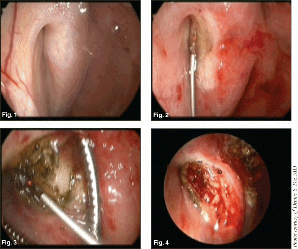
The Procedure

Eustachian tuboplasty is a nasal or transoral endoscopic outpatient procedure requiring general anesthesia. To date, it has been used only in adults, said Dr. Poe. A laser or microdebrider is used to strip away hypertrophic mucosa and cartilage on the posterior eustachian tube cushion and into an area called the valve to clear obstruction.

According to a recent paper describing microdebridement in 20 patients (Otolaryngol Head Neck Surg 2007;136:422-7), Dr. Poe and Dr. Metson describe the valve as a 0.5-cm-long region where mucosal surfaces from the anterior and posterior cushions are seen to appose during tubal closure and dilate to the open position with normal tubal function.

During surgery, physicians take special care to steer clear of injury to the mucosa on the anterior cushion to avoid postoperative scarring and stenosis of the eustachian tube orifice.

Pages: 1 2 3 4 5 6 | Single Page

Filed Under: Everyday Ethics, Head and Neck, Medical Education, Otology/Neurotology, Practice Management Tagged With: Otology, Patulous eustacian tube, tuboplastyIssue: July 2009

You Might Also Like:

  • Eustachian Tube Scores Effective Diagnostic Tools for Chronic Obstructive Eustachian Tube Dysfunction
  • New Developments in the Management of Eustachian Tube Dysfunction
  • Patulous Eustachian Tube Surgical Treatment Safe, Effective
  • Intranasal Corticosteroids Treatment Shown Ineffective for Chronic Eustachian Tube Dysfunction

Leave a Reply Cancel reply

Your email address will not be published. Required fields are marked *

The Triological SocietyENTtoday is a publication of The Triological Society.

Polls

Would you choose a concierge physician as your PCP?

View Results

Loading ... Loading ...
  • Polls Archive

Top Articles for Residents

  • Applications Open for Resident Members of ENTtoday Edit Board
  • How To Provide Helpful Feedback To Residents
  • Call for Resident Bowl Questions
  • New Standardized Otolaryngology Curriculum Launching July 1 Should Be Valuable Resource For Physicians Around The World
  • Do Training Programs Give Otolaryngology Residents the Necessary Tools to Do Productive Research?
  • Popular this Week
  • Most Popular
  • Most Recent
    • A Journey Through Pay Inequity: A Physician’s Firsthand Account

    • The Dramatic Rise in Tongue Tie and Lip Tie Treatment

    • Otolaryngologists Are Still Debating the Effectiveness of Tongue Tie Treatment

    • Is Middle Ear Pressure Affected by Continuous Positive Airway Pressure Use?

    • Rating Laryngopharyngeal Reflux Severity: How Do Two Common Instruments Compare?

    • The Dramatic Rise in Tongue Tie and Lip Tie Treatment

    • Rating Laryngopharyngeal Reflux Severity: How Do Two Common Instruments Compare?

    • Is Middle Ear Pressure Affected by Continuous Positive Airway Pressure Use?

    • Otolaryngologists Are Still Debating the Effectiveness of Tongue Tie Treatment

    • Complications for When Physicians Change a Maiden Name

    • Excitement Around Gene Therapy for Hearing Restoration
    • “Small” Acts of Kindness
    • How To: Endoscopic Total Maxillectomy Without Facial Skin Incision
    • Science Communities Must Speak Out When Policies Threaten Health and Safety
    • Observation Most Cost-Effective in Addressing AECRS in Absence of Bacterial Infection

Follow Us

  • Contact Us
  • About Us
  • Advertise
  • The Triological Society
  • The Laryngoscope
  • Laryngoscope Investigative Otolaryngology
  • Privacy Policy
  • Terms of Use
  • Cookies

Wiley

Copyright © 2025 by John Wiley & Sons, Inc. All rights reserved, including rights for text and data mining and training of artificial technologies or similar technologies. ISSN 1559-4939