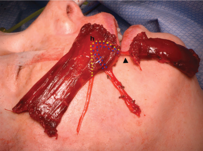
Fig. 1. Intraoperative photograph of harvested ipsilateral flap demonstrating orientation of upper and lower lip paddles following 180° in-plane rotation. In this iteration, the nerve and vascular pedicle are reflected deep to the flap for coaptation with the ipsilateral nerve-to-masseter muscle and anastomosis with the facial vessels. The approximate location of the hilum (h) and course of the neurovascular pedicle (dotted lines: yellow—nerve-to-gracilis muscle, blue—venae comitans, red—artery) on the deep aspect of the flap is demonstrated, together with the continuation of the neurovascular pedicle (arrowhead) to the lower lip paddle.