• Home
  • Practice Focus
    • Facial Plastic/Reconstructive
    • Head and Neck
    • Laryngology
    • Otology/Neurotology
    • Pediatric
    • Rhinology
    • Sleep Medicine
    • How I Do It
    • TRIO Best Practices
  • Business of Medicine
    • Health Policy
    • Legal Matters
    • Practice Management
    • Tech Talk
    • AI
  • Literature Reviews
    • Facial Plastic/Reconstructive
    • Head and Neck
    • Laryngology
    • Otology/Neurotology
    • Pediatric
    • Rhinology
    • Sleep Medicine
  • Career
    • Medical Education
    • Professional Development
    • Resident Focus
  • ENT Perspectives
    • ENT Expressions
    • Everyday Ethics
    • From TRIO
    • The Great Debate
    • Letter From the Editor
    • Rx: Wellness
    • The Voice
    • Viewpoint
  • TRIO Resources
    • Triological Society
    • The Laryngoscope
    • Laryngoscope Investigative Otolaryngology
    • TRIO Combined Sections Meetings
    • COSM
    • Related Otolaryngology Events
  • Search

Single-Port Endoscopic Removal of Forehead Osteoma: An Otolaryngologist’s Procedure

by Kachorn Seresirikachorn, MD, PhD, Lu Hui Png, MD, and Richard J. Harvey, MD, PhD • January 22, 2024

  • Tweet
  • Click to email a link to a friend (Opens in new window) Email
Print-Friendly Version

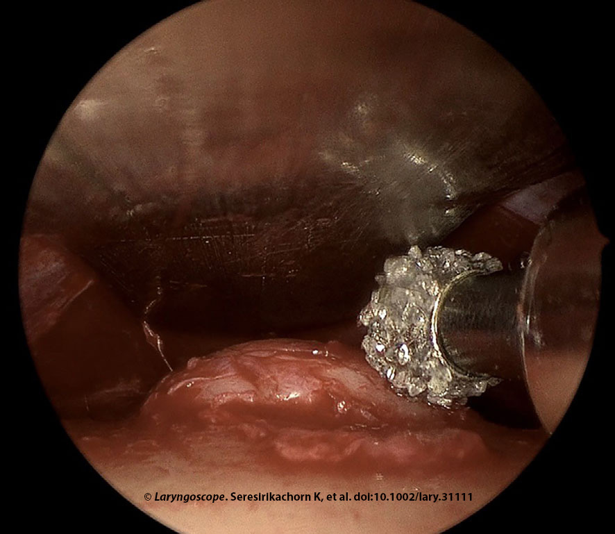
Figure 1. Endoscopic forehead osteoma removal was done in a 49-year-old female with an 11 mm left forehead osteoma. Drilling is shown here.

The forehead osteoma removal was performed with a 4-mm 15° diamond burr at 30,000 revolutions per minute, along with irrigation and suction system under 0° endoscope visualization. Irrigation and suction were critical for drilling to reduce heat and clean the surgical field. The surgeon held the 0° endoscope above the drill, whereas the assistant held the malleable retractor. The uneven frontal bone surface was recontoured with drilling. The ethmoid curette was used to smooth any minor irregularities after drilling (Figure 1). Finally, irrigation with normal saline was performed to clean the operative field and ensure that all bone debris was removed. The wound was closed using absorbable sutures (5-0 coated Vicryl and 5-0 Plain Gut). The forehead was compressed with an elastic bandage for three to five days postoperative.

You Might Also Like

  • How To: Hidden Port Approach to Endoscopic Pericranial Scalp Flap for Anterior Skull Base Reconstruction
  • Potential Advantage Using Robotic Thyroidectomy via Posterior Neck Approach with the DaVinci Single Port System
  • What Is the Optimal Timing for Dividing a Forehead Flap?
  • How To: Transoral Endoscopic Vestibular Approach to the Sistrunk Procedure
Explore This Issue
January 2024

Postoperative Care

Patients were managed via day surgery. Amoxicillin/clavulanic acid was given for 10 days, and prednisone was given at 25 mg daily for seven days to reduce swelling.

RESULTS

Nine patients (48.9 ± 10.1 years, 100% female) were assessed. The osteoma diameter was 11.7 ± 3.5 mm. Multiple osteomas were present in 29%. All patients had complete removal of forehead osteoma (100% [95% CI: 66.4%–100%]) and success in restoration of contour (100% [95% CI: 66.4%–100%]). The follow-up was 24.8 ± 19.1 months. There were no early (0% [95% CI: 0%–33.6%]) or late (0% [95% CI: 0%–33.6%]) surgical morbidities.

Similar outcomes have been reported with complete removal and excellent cosmesis. The technique described utilizes familiar drills and endoscopic equipment for the otolaryngologist. The subperiosteal dissection and osteoma removal steps were performed via endoscopic visualization. Although performing osteoma removal without endoscope visualization using osteotome and hammer instrumentation has been described (Formos J Surg. 2015. doi:10.1016/j.fjs.2015.04.001), the endoscope provides precise drilling and limits soft tissue dissection. Additionally, this skill set is very familiar to otolaryngologists.

The risk of supraorbital or supratrochlear nerve injury, and subsequent paresthesia, are often minimized by the subperiosteal approach, the high incision, and small port access. In our series, the entire dissection and tumor removal were performed via a single remote port. Multiple port approaches for endoscopic removal of forehead osteoma, attributed to the complexity arising from an excessively narrow inter-instrument angle that impedes manipulation, have been described in the literature but are not required. Otolaryngologists are familiar with endoscopic visualization via a single port, similar to that used for endoscopic sinus or ear surgery.

Pages: 1 2 3 | Single Page

Filed Under: Head and Neck, How I Do It, Practice Focus Tagged With: endoscopic surgery, osteomaIssue: January 2024

You Might Also Like:

  • How To: Hidden Port Approach to Endoscopic Pericranial Scalp Flap for Anterior Skull Base Reconstruction
  • Potential Advantage Using Robotic Thyroidectomy via Posterior Neck Approach with the DaVinci Single Port System
  • What Is the Optimal Timing for Dividing a Forehead Flap?
  • How To: Transoral Endoscopic Vestibular Approach to the Sistrunk Procedure

The Triological SocietyENTtoday is a publication of The Triological Society.

Polls

Would you choose a concierge physician as your PCP?

View Results

Loading ... Loading ...
  • Polls Archive

Top Articles for Residents

  • Applications Open for Resident Members of ENTtoday Edit Board
  • How To Provide Helpful Feedback To Residents
  • Call for Resident Bowl Questions
  • New Standardized Otolaryngology Curriculum Launching July 1 Should Be Valuable Resource For Physicians Around The World
  • Do Training Programs Give Otolaryngology Residents the Necessary Tools to Do Productive Research?
  • Popular this Week
  • Most Popular
  • Most Recent
    • The Dramatic Rise in Tongue Tie and Lip Tie Treatment

    • Shifting the Treatment Goalpost Toward Medical Management of Recurrent Respiratory Papillomatosis

    • Otolaryngologists Are Still Debating the Effectiveness of Tongue Tie Treatment

    • Rating Laryngopharyngeal Reflux Severity: How Do Two Common Instruments Compare?

    • A Journey Through Pay Inequity: A Physician’s Firsthand Account

    • The Dramatic Rise in Tongue Tie and Lip Tie Treatment

    • Rating Laryngopharyngeal Reflux Severity: How Do Two Common Instruments Compare?

    • Is Middle Ear Pressure Affected by Continuous Positive Airway Pressure Use?

    • Otolaryngologists Are Still Debating the Effectiveness of Tongue Tie Treatment

    • Complications for When Physicians Change a Maiden Name

    • Excitement Around Gene Therapy for Hearing Restoration
    • “Small” Acts of Kindness
    • How To: Endoscopic Total Maxillectomy Without Facial Skin Incision
    • Science Communities Must Speak Out When Policies Threaten Health and Safety
    • Observation Most Cost-Effective in Addressing AECRS in Absence of Bacterial Infection

Follow Us

  • Contact Us
  • About Us
  • Advertise
  • The Triological Society
  • The Laryngoscope
  • Laryngoscope Investigative Otolaryngology
  • Privacy Policy
  • Terms of Use
  • Cookies

Wiley

Copyright © 2025 by John Wiley & Sons, Inc. All rights reserved, including rights for text and data mining and training of artificial technologies or similar technologies. ISSN 1559-4939