INTRODUCTION
Forehead osteoma is a benign tumor lesion, although mostly asymptomatic, creating significant cosmetic concerns for the patient. Traditionally, either a direct open approach with a forehead incision along the relaxed free tension line or a large coronal incision in the hair-bearing skin is made to remove forehead osteoma. However, an inevitable scar is the main issue for a direct open approach (J Craniofac Surg. 2006. doi:10.1097/00001665-200605000-00007). Additionally, there is the risk of temporary or permanent scalp paresthesia from the supraorbital or supratrochlear nerve injury.
Explore This Issue
January 2024Endoscope-assisted removal of forehead osteoma has been described, but it is not clear to many patients which surgical specialty has the appropriate training for this approach (J Craniofac Surg. 2006. doi:10.1097/00001665-200605000-00007; J Craniofac Surg. 1995. doi:10.1097/00001665-199511000-00021; Formos J Surg. 2015. doi:10.1097/SCS.0000000000005185; Ann Plast Surg. 2008. doi:10.1097/SAP.0b013e31816d829a; Ann Chir Plast Esthet. 2020. doi:10.1016/j.anplas.2019.04.004). Referrals to plastic surgeons, neurosurgeons, and general surgeons are common, but otolaryngologists have the core skill set required for this procedure. The excellent visualization from the endoscope creates success in complete removal and recontouring of the forehead without nerve injury. Moreover, the hidden incision in the frontal hairline gives excellent cosmetic outcomes.
Here, we describe our single-port endoscopic removal of forehead osteoma technique and the associated outcomes.
METHOD
A retrospective review of consecutive adult patients with forehead osteoma managed with single-port endoscopic removal of forehead osteoma was performed. Success in the primary outcomes was measured by the complete removal of the osteoma, which could not be observed by either the patient or the surgeon, and the restoration of contour without irregularities in the forehead, which also could not be seen or palpated by the patient and the surgeon. Secondary outcomes were surgical morbidity, defined as early (<90 days) or late (>90 days). Early morbidity included periorbital swelling/bruising, infection, temporary scalp paresthesia, forehead hematoma, and pain requiring additional analgesia. Late morbidity included permanent scalp paresthesia, irregularity of forehead contour, and visible surgical scarring.
Preparation
The operation was performed under general anesthesia. The surgeon operated in the sitting position at the top of the operating bed. The endoscope monitor was placed above the patient in front of the surgeon. The patient’s head was in a neutral anatomic position. The boundary of frontal osteoma was marked. The hair was divided by bush up and down above the frontal hairline without a haircut. The hair division line was perpendicular to the osteoma. The “deckled” hairline incision of 1–2 cm was drawn in the hair division line. The incision, tumor site, and endoscopic instruments’ access path were infiltrated with 1% ropivacaine and 1:100,000 adrenaline.
Surgical Technique
The deckled hairline incision was made with the 15° ophthalmic knife. Needle-point diathermy coagulation occurred to the galeal layer, setting 12. Under retraction, the surgeon blunt dissected, vertically through the galea, to the subperiosteal plane down to the subperiosteal layer. Then, a 4-mm 0° endoscope was inserted under the subperiosteal plane. The subperiosteal dissection was performed down to the osteoma’s cephalic and lateral border, extending laterally to ensure full visualization. Dissection inferior and below the border of the osteoma was facilitated with an ethmoid curette.
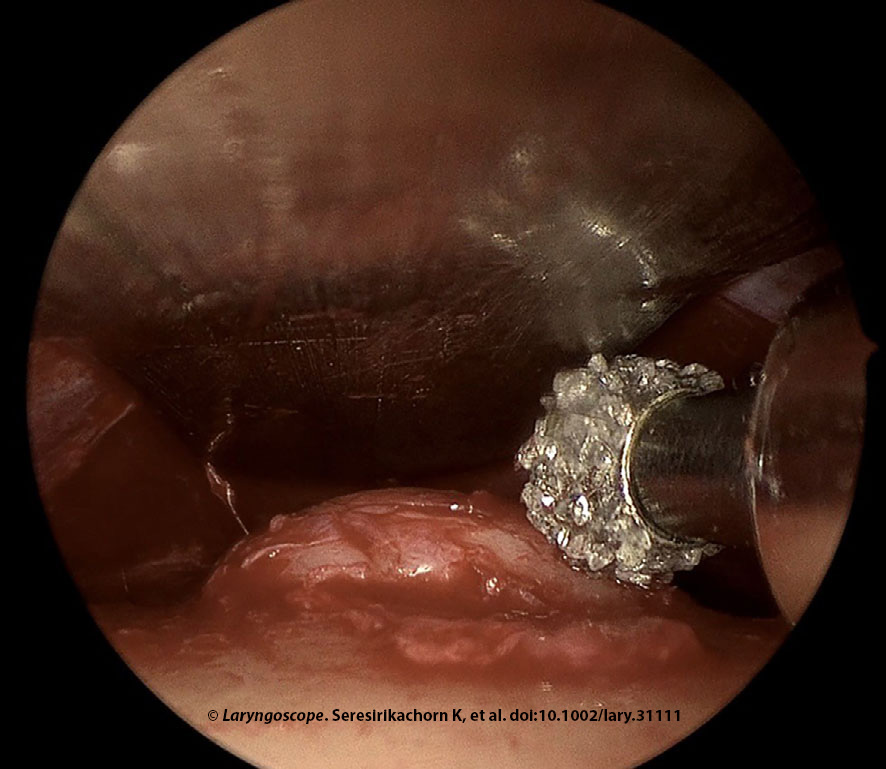
Figure 1. Endoscopic forehead osteoma removal was done in a 49-year-old female with an 11 mm left forehead osteoma. Drilling is shown here.
The forehead osteoma removal was performed with a 4-mm 15° diamond burr at 30,000 revolutions per minute, along with irrigation and suction system under 0° endoscope visualization. Irrigation and suction were critical for drilling to reduce heat and clean the surgical field. The surgeon held the 0° endoscope above the drill, whereas the assistant held the malleable retractor. The uneven frontal bone surface was recontoured with drilling. The ethmoid curette was used to smooth any minor irregularities after drilling (Figure 1). Finally, irrigation with normal saline was performed to clean the operative field and ensure that all bone debris was removed. The wound was closed using absorbable sutures (5-0 coated Vicryl and 5-0 Plain Gut). The forehead was compressed with an elastic bandage for three to five days postoperative.
Postoperative Care
Patients were managed via day surgery. Amoxicillin/clavulanic acid was given for 10 days, and prednisone was given at 25 mg daily for seven days to reduce swelling.
RESULTS
Nine patients (48.9 ± 10.1 years, 100% female) were assessed. The osteoma diameter was 11.7 ± 3.5 mm. Multiple osteomas were present in 29%. All patients had complete removal of forehead osteoma (100% [95% CI: 66.4%–100%]) and success in restoration of contour (100% [95% CI: 66.4%–100%]). The follow-up was 24.8 ± 19.1 months. There were no early (0% [95% CI: 0%–33.6%]) or late (0% [95% CI: 0%–33.6%]) surgical morbidities.
Similar outcomes have been reported with complete removal and excellent cosmesis. The technique described utilizes familiar drills and endoscopic equipment for the otolaryngologist. The subperiosteal dissection and osteoma removal steps were performed via endoscopic visualization. Although performing osteoma removal without endoscope visualization using osteotome and hammer instrumentation has been described (Formos J Surg. 2015. doi:10.1016/j.fjs.2015.04.001), the endoscope provides precise drilling and limits soft tissue dissection. Additionally, this skill set is very familiar to otolaryngologists.
The risk of supraorbital or supratrochlear nerve injury, and subsequent paresthesia, are often minimized by the subperiosteal approach, the high incision, and small port access. In our series, the entire dissection and tumor removal were performed via a single remote port. Multiple port approaches for endoscopic removal of forehead osteoma, attributed to the complexity arising from an excessively narrow inter-instrument angle that impedes manipulation, have been described in the literature but are not required. Otolaryngologists are familiar with endoscopic visualization via a single port, similar to that used for endoscopic sinus or ear surgery.