• Home
  • Practice Focus
    • Facial Plastic/Reconstructive
    • Head and Neck
    • Laryngology
    • Otology/Neurotology
    • Pediatric
    • Rhinology
    • Sleep Medicine
    • How I Do It
    • TRIO Best Practices
  • Business of Medicine
    • Health Policy
    • Legal Matters
    • Practice Management
    • Tech Talk
    • AI
  • Literature Reviews
    • Facial Plastic/Reconstructive
    • Head and Neck
    • Laryngology
    • Otology/Neurotology
    • Pediatric
    • Rhinology
    • Sleep Medicine
  • Career
    • Medical Education
    • Professional Development
    • Resident Focus
  • ENT Perspectives
    • ENT Expressions
    • Everyday Ethics
    • From TRIO
    • The Great Debate
    • Letter From the Editor
    • Rx: Wellness
    • The Voice
    • Viewpoint
  • TRIO Resources
    • Triological Society
    • The Laryngoscope
    • Laryngoscope Investigative Otolaryngology
    • TRIO Combined Sections Meetings
    • COSM
    • Related Otolaryngology Events
  • Search

Studies Examine Semicircular Canal Dehiscence, Stapedectomy Prostheses.

by Pippa Wysong • September 1, 2008

  • Tweet
  • Click to email a link to a friend (Opens in new window) Email
Print-Friendly Version

Be careful not to be too quick to say that a patient’s problems are due to canal dehiscence. A study using CT to determine the prevalence of superior and posterior semicircular canal dehiscence in a patient population in Boston calculated the prevalence of superior canal dehiscence; although the result was similar to those from previous investigations (about 9%), this study shows there is a twist-the rate of posterior canal dehiscence may be lower than previously reported.

You Might Also Like

  • Is There a “Best” Stapedectomy Prosthesis?
  • How to: Positioning for Middle Cranial Fossa Repair of Superior Semicircular Canal Dehiscence
  • Certain Approaches to Superior Semicircular Canal Dehiscence Repair Are Effective for Favorable Hearing Outcomes for Patients
  • Dehiscence Size in SSCD Correlates with Air-Bone Gap Size
Explore This Issue
September 2008

Previously published studies were in general, large sample size populations but they did not employ sub-millimeter technique. We aren’t aware of any investigations employing sub-millimeter technique in evaluating the posterior semicircular canal, said Rohini Nadgir, MD, Assistant Professor of Radiology at the Boston University School of Medicine, who spoke at a Triological Society session at the Combined Otolaryngology Spring Meeting.

In this retrospective study, researchers reviewed CT scans from patients who had undergone temporal bone CT scans for a variety of clinical indications between July 2005 and March 2007. Scans were done using a sub-millimeter technique to evaluate the integrity of the superior and posterior semicircular canals. The scans were performed on a 64 detector row CT scanner using 0.625-mm slice thickness, in bone plus algorithm without contrast. A review was also done of 0.3-mm interval reconstructions in the coronal and sagittal planes.

The images were reviewed by two neuro-radiologists who rated the superior and posterior canals as normal, thin, or dehiscent. Clinically relevant data were reviewed by an otolaryngologic surgeon who was blinded to the interpretations of the CT. Radiographic results were correlated to the clinical findings.
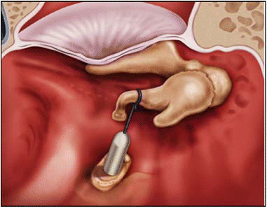
Figure. The Nitinol prosthesis avoids contact between the tip and the piston, increasing contact with the incus and avoiding an oval crimp.Courtesy of Gyrus ACMI.

click for large version
Figure. The Nitinol prosthesis avoids contact between the tip and the piston, increasing contact with the incus and avoiding an oval crimp.Courtesy of Gyrus ACMI.

Images from 454 temporal bones from 226 patients were reviewed. Of these, 19 (9%) patients were found to have dehiscence of the superior semicircular canal. Intriguingly, of these patients, none had supportive clinical evidence of the syndrome, she said. Two patients were identified as having posterior canal dehiscence, and they didn’t have clinical signs either.

Ironically, through the chart review, five patients had an initial clinical suspicion of canal dehiscence, but none had supportive radiographic evidence. The patients were found to have other explanations for their symptoms, including benign positional vertigo, endolymphatic hydrops, cholesteatoma, and otosclerosis.

Overall, the findings confirmed the same rate of superior canal dehiscence as previous studies; however, the rate of posterior canal dehiscence was much lower than published rates.

Pages: 1 2 3 | Single Page

Filed Under: Departments, Medical Education, Otology/Neurotology, Practice Focus, Tech Talk Tagged With: CT, Otology, outcomes, prosthesis, research, stapedectomy, technologyIssue: September 2008

You Might Also Like:

  • Is There a “Best” Stapedectomy Prosthesis?
  • How to: Positioning for Middle Cranial Fossa Repair of Superior Semicircular Canal Dehiscence
  • Certain Approaches to Superior Semicircular Canal Dehiscence Repair Are Effective for Favorable Hearing Outcomes for Patients
  • Dehiscence Size in SSCD Correlates with Air-Bone Gap Size

Leave a Reply Cancel reply

Your email address will not be published. Required fields are marked *

The Triological SocietyENTtoday is a publication of The Triological Society.

Polls

Would you choose a concierge physician as your PCP?

View Results

Loading ... Loading ...
  • Polls Archive

Top Articles for Residents

  • Applications Open for Resident Members of ENTtoday Edit Board
  • How To Provide Helpful Feedback To Residents
  • Call for Resident Bowl Questions
  • New Standardized Otolaryngology Curriculum Launching July 1 Should Be Valuable Resource For Physicians Around The World
  • Do Training Programs Give Otolaryngology Residents the Necessary Tools to Do Productive Research?
  • Popular this Week
  • Most Popular
  • Most Recent
    • A Journey Through Pay Inequity: A Physician’s Firsthand Account

    • The Dramatic Rise in Tongue Tie and Lip Tie Treatment

    • Otolaryngologists Are Still Debating the Effectiveness of Tongue Tie Treatment

    • Is Middle Ear Pressure Affected by Continuous Positive Airway Pressure Use?

    • Rating Laryngopharyngeal Reflux Severity: How Do Two Common Instruments Compare?

    • The Dramatic Rise in Tongue Tie and Lip Tie Treatment

    • Rating Laryngopharyngeal Reflux Severity: How Do Two Common Instruments Compare?

    • Is Middle Ear Pressure Affected by Continuous Positive Airway Pressure Use?

    • Otolaryngologists Are Still Debating the Effectiveness of Tongue Tie Treatment

    • Complications for When Physicians Change a Maiden Name

    • Excitement Around Gene Therapy for Hearing Restoration
    • “Small” Acts of Kindness
    • How To: Endoscopic Total Maxillectomy Without Facial Skin Incision
    • Science Communities Must Speak Out When Policies Threaten Health and Safety
    • Observation Most Cost-Effective in Addressing AECRS in Absence of Bacterial Infection

Follow Us

  • Contact Us
  • About Us
  • Advertise
  • The Triological Society
  • The Laryngoscope
  • Laryngoscope Investigative Otolaryngology
  • Privacy Policy
  • Terms of Use
  • Cookies

Wiley

Copyright © 2025 by John Wiley & Sons, Inc. All rights reserved, including rights for text and data mining and training of artificial technologies or similar technologies. ISSN 1559-4939