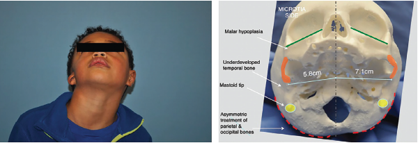
Figure 1. Left: preoperative image demonstrating degree of hemifacial microsomia highlighted by deficient mastoid and asymmetric midface and mandible. Right: 3D-printed skull highlighting these changes at the bony level.
Figure 1. Left: preoperative image demonstrating degree of hemifacial microsomia highlighted by deficient mastoid and asymmetric midface and mandible. Right: 3D-printed skull highlighting these changes at the bony level.