• Home
  • Practice Focus
    • Facial Plastic/Reconstructive
    • Head and Neck
    • Laryngology
    • Otology/Neurotology
    • Pediatric
    • Rhinology
    • Sleep Medicine
    • How I Do It
    • TRIO Best Practices
  • Business of Medicine
    • Health Policy
    • Legal Matters
    • Practice Management
    • Tech Talk
    • AI
  • Literature Reviews
    • Facial Plastic/Reconstructive
    • Head and Neck
    • Laryngology
    • Otology/Neurotology
    • Pediatric
    • Rhinology
    • Sleep Medicine
  • Career
    • Medical Education
    • Professional Development
    • Resident Focus
  • ENT Perspectives
    • ENT Expressions
    • Everyday Ethics
    • From TRIO
    • The Great Debate
    • Letter From the Editor
    • Rx: Wellness
    • The Voice
    • Viewpoint
  • TRIO Resources
    • Triological Society
    • The Laryngoscope
    • Laryngoscope Investigative Otolaryngology
    • TRIO Combined Sections Meetings
    • COSM
    • Related Otolaryngology Events
  • Search

Optical Coherence Tomography Offers Noninvasive Way to Visualize Tissues of the Larynx: This emerging technology could improve testing for laryngeal cancers

by Coriene E. Hannapel • September 1, 2006

  • Tweet
  • Click to email a link to a friend (Opens in new window) Email
Print-Friendly Version

OCT is not as good as microscopy, but you see much deeper in the tissue, on the order of about a millimeter, with very high resolution. Current systems in our laboratory are approaching one micron resolution, Dr. Wong added.

You Might Also Like

  • Emerging Use of Optical Coherence Tomography in Otolaryngology
  • Dynamic Optical Contrast Imaging Aids Parathyroid Gland Localization in Ex Vivo Specimens
  • DRS/FS Help Identify Nerves from Adjacent Tissues
  • Technology Improves Management of Frontal Sinus Fractures
Explore This Issue
September 2006

Future

This technology will likely follow the same adoption and usage dynamics as CT, ultrasound, and MRI, Dr. Wong said.

Figure. The prototype head and neck optical coherence tomography (OTC) imaging device, pictured here, must be delivered through an endoscope to an anesthetized patient, but researchers would like to develop a version that can be used in the office while the patient is awake.

click for large version
Figure. The prototype head and neck optical coherence tomography (OTC) imaging device, pictured here, must be delivered through an endoscope to an anesthetized patient, but researchers would like to develop a version that can be used in the office while the patient is awake.

The OCT procedure will require training, Dr. Wong added. I think that this is the type of technology that is probably going to make its way into residency training programs first, but I think that clinicians who want to use OCT in their offices are probably going to take some CME courses.

With new imaging modalities, the line between surgeon, pathologist, and radiologist is blurring, Dr. Wong noted. OCT technology is certainly going to involve pathologists, but a pathologist is not in the operating room with the patient-there are other technologies that are being developed where surgeons are really going to have to understand how to do imaging. This is already happening in other applications-surgeons know how to interpret ultrasound so they can use the information intraoperatively or in the office or clinic. So there is certainly a precedent for that, Dr. Wong added.

More work still needs to be done in terms of instrumentation design and technology improvement, Dr. Wong noted. The capabilities of OCT need to be improved in terms of acquisition time speed and resolution. This is an active area of research by investigators in several institutions in optical sciences in the US, Europe, and Asia.

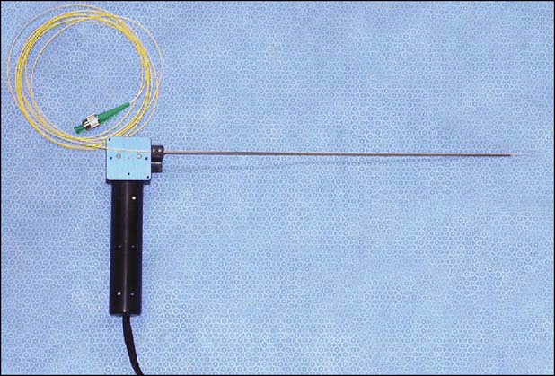
We are trying to identify and characterize both normal and pathological structures and we are still in the early stages in terms of optimizing the equipment in terms of ergonomics, of being able to use it in a non-invasive manner, and in terms of miniaturization, Dr. Armstrong said. Research work is also focusing on improving the software and hardware so that it can be easily used in a clinical situation and be adaptable to the clinician.

Research systems at UC-Irvine could be taken into clinical implementation in the next two years, the researchers predict.

Pages: 1 2 3 4 5 | Single Page

Filed Under: Departments, Laryngology, Medical Education, Practice Focus, Tech Talk Tagged With: CT, Imaging, research, technology, testing, training, vocal foldsIssue: September 2006

You Might Also Like:

  • Emerging Use of Optical Coherence Tomography in Otolaryngology
  • Dynamic Optical Contrast Imaging Aids Parathyroid Gland Localization in Ex Vivo Specimens
  • DRS/FS Help Identify Nerves from Adjacent Tissues
  • Technology Improves Management of Frontal Sinus Fractures

Leave a Reply Cancel reply

Your email address will not be published. Required fields are marked *

The Triological SocietyENTtoday is a publication of The Triological Society.

Polls

Have you invented or patented something that betters the field of otolaryngology?

View Results

Loading ... Loading ...
  • Polls Archive

Top Articles for Residents

  • Applications Open for Resident Members of ENTtoday Edit Board
  • How To Provide Helpful Feedback To Residents
  • Call for Resident Bowl Questions
  • New Standardized Otolaryngology Curriculum Launching July 1 Should Be Valuable Resource For Physicians Around The World
  • Do Training Programs Give Otolaryngology Residents the Necessary Tools to Do Productive Research?
  • Popular this Week
  • Most Popular
  • Most Recent
    • The Dramatic Rise in Tongue Tie and Lip Tie Treatment

    • Otolaryngologists as Entrepreneurs: Transforming Patient Care And Practice

    • Otolaryngologists Are Still Debating the Effectiveness of Tongue Tie Treatment

    • Continued Discussion And Engagement Are Essential To How Otolaryngologists Are Championing DEI Initiatives In Medicine

    • Rating Laryngopharyngeal Reflux Severity: How Do Two Common Instruments Compare?

    • The Dramatic Rise in Tongue Tie and Lip Tie Treatment

    • Rating Laryngopharyngeal Reflux Severity: How Do Two Common Instruments Compare?

    • Is Middle Ear Pressure Affected by Continuous Positive Airway Pressure Use?

    • Otolaryngologists Are Still Debating the Effectiveness of Tongue Tie Treatment

    • Complications for When Physicians Change a Maiden Name

    • Leaky Pipes—Time to Focus on Our Foundations
    • You Are Among Friends: The Value Of Being In A Group
    • How To: Full Endoscopic Procedures of Total Parotidectomy
    • How To: Does Intralesional Steroid Injection Effectively Mitigate Vocal Fold Scarring in a Rabbit Model?
    • What Is the Optimal Anticoagulation in HGNS Surgery in Patients with High-Risk Cardiac Comorbidities?

Follow Us

  • Contact Us
  • About Us
  • Advertise
  • The Triological Society
  • The Laryngoscope
  • Laryngoscope Investigative Otolaryngology
  • Privacy Policy
  • Terms of Use
  • Cookies

Wiley

Copyright © 2025 by John Wiley & Sons, Inc. All rights reserved, including rights for text and data mining and training of artificial technologies or similar technologies. ISSN 1559-4939